Book - Vertebrate Zoology (1928) 19
Vertebrate Zoology G. R. De Beer (1928)
| Historic Disclaimer - information about historic embryology pages |
|---|
| Pages where the terms "Historic" (textbooks, papers, people, recommendations) appear on this site, and sections within pages where this disclaimer appears, indicate that the content and scientific understanding are specific to the time of publication. This means that while some scientific descriptions are still accurate, the terminology and interpretation of the developmental mechanisms reflect the understanding at the time of original publication and those of the preceding periods, these terms, interpretations and recommendations may not reflect our current scientific understanding. (More? Embryology History | Historic Embryology Papers) |
Chapter XIX The Coelom And Mesoderm
In Amphioxus and all Craniates the most dorsal mesoderm is segmented into somites. These each contain a portion of coelomic cavity called myoccel, which persists in Amphioxus, but becomes obliterated in higher forms. The median wall of the myocoel is thickened and produces the myotome : a plate of muscle with striated fibres, innervated by somatic efferent fibres (voluntary) through the ventral nerve-roots. The outer layer of coelomic epithelium lateral to the myocoel gives rise to the dermatome or cutis-layer, beneath the skin. On the median side, the myotome also produces the sclerotome. In Amphioxus this is in the form of a hollow outgrowth, but in higher forms it is composed of mesenchyme. It gives rise in Craniates to the basidorsal and basiventral elements which go to make up the vertebral column.
The dorsal segmented portion of the mesoderm is known as the vertebral plate. The more ventral portion of the mesoderm arises segmentally in Amphioxus, each segment separated from the ones in front and behind by septa. These septa, however, break down, and the ventral coelomic cavity or splanchnocoel is continuous from end to end of the animal. This condition arises from the first in the Craniates, where the mesoderm in this region, known as the lateral plate, is not segmented. The outer wall of the splanchnocoel becomes applied to the body- wall, and the inner wall covers the gut- wall. The separation between right and left splanchnocoel usually breaks down ventrally, but persists dorsally as the mesentery which suspends the gut. The muscles which the coelomic epithelium of the splanchnocoel produces are smooth, involuntary, and innervated by the autonomic nervous system, except for those which are situated in the anterior region of the body, in connexion with the gill-slits. The gill-slits pierce through from the gut to the outside in the region of the lateral plate ; between the gill-slits, in the visceral arches, the lateral-plate mesoderm gives rise to the muscles which move the arches, including the jaws. These muscles are striated and voluntary, but they are not myotomic, and they are innervated by visceral efferent fibres through the dorsal roots of the cranial nerves.
Between the myoccels and the splanchnocoels there are typically little hollow stalks, through which at early stages the cavities of the latter can communicate with those of the former. They are segmental in arrangement. In Amphioxus, these regions of the coelom represent the future gonads, and are called the gonotomes with their cavities the gonocoels. In the Craniates, they are called the nephrotomes (or intermediate cell-masses) ; the cavities (communications between the myocoels and the splanchnocoel) are the nephrocoels, and they give rise to the tubules of the kidneys and associated structures, eventually losing connexion both with myocoels and splanch- nocoel.
In Amphioxus the splanchnocoel is continuous from end to end of the body as in the Ammocoete, for the transverse septum in which the ductus Cuvieri crosses over from the body- wall to the gut- wall, is not large. In Selachians, the transverse septum separates an anterior pericardial cavity from a posterior peritoneal or perivisceral cavity, leaving only very small communications between them in the form of the pericardio-peritoneal canals. In higher forms the separation between pericardial and perivisceral cavities is complete. Beginning in the Dipnoi, the pericardium becomes thin- walled and projects backwards into the perivisceral cavity.
All viscera are morphologically outside the coelomic cavity and only suspended in it by a bag of coelomic epithelium which forms a double membrane or mesentery. So the gut is suspended by the dorsal mesentery from the roof of the perivisceral cavity, and between the two membranes composing it there pass the arteries from the dorsal aorta to the gut. The gut and liver are connected by the lesser omentum, through which the bile-duct runs from the liver to the anterior portion of the intestine. The lungs in amphibia are of course covered over by coelomic epithelium (pleura) which is continuous with the ordinary lining of the perivisceral cavity round the stalk of the lungs. In some reptiles, the coelomic epithelium covering the lung is also attached to the roof of the perivisceral cavity forming the accessory mesentery, and attached to the liver below by the pulmo-hepatic ligaments. On each side of the dorsal mesentery therefore there is a recess, the pulmo-hepatic recess, bounded on the median side by the dorsal mesentery and stomach, laterally by the accessory mesentery and pulmo-hepatic ligament, and below by the liver. Owing to the curvature of the stomach and the return of the anterior portion of the intestine to form the loop of the duodenum, the pulmo- hepatic recess of the right side comes to form a pocket, the
Fig. 125. Transverse section through the trunk of an embryo of Lacerta, showing the relations of the coelom.da, dorsal aorta ; dm, dorsal mesentery ; fl, I, liver ; In, lung ; lo, lesser omentum ; n t phi, pulmo-hepatic ligament ; phr, pulmo- am, accessory mesentery ; falciform ligament ; g, gut ; notochord ; nc, nerve-cord ; hepatic recess.
Fig. 126. Transverse section through the trunk of a bird showing the relations of the coelom. as, air-sac ; os, oblique septum ; pc, pleural cavity ; v, vertebra ; other letters as Fig. 125.
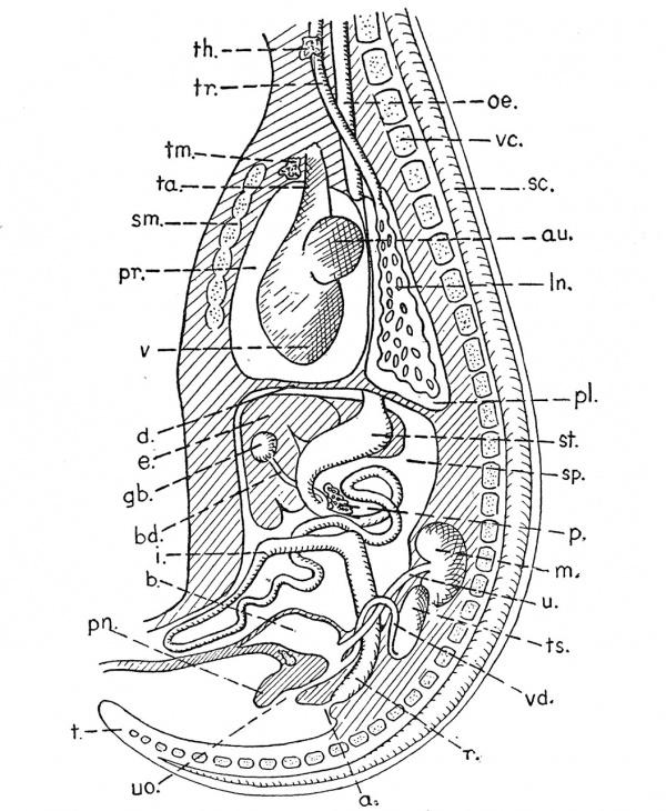
In many mammals, the dorsal mesentery supporting the stomach from the roof of the perivisceral cavity, becomes drawn out into a double sheet of coelomic epithelium which overlaps the transverse colon of the large intestine on the ventral side. Eventually this sheet may fuse with the mesentery suspending the large intestine (mesocolon). This extension, which is called the great omentum, brings about an increase in size of the omental sac, on the wall of which fat is often deposited.
Fig. 127. Longitudinal section through the trunk of a mammalian embryo, showing the relations of the coelom and viscera. a, anus ; au, auricle of heart ; b, bladder (continuous with allantois) ; bd, bile-duct ; d, diaphragm ; e, liver ; gb, gall-bladder ; i, intestine ; In, lung ; m, metanephric kidney ; oe, oesophagus ; p, pancreas ; pi, pleural coelomic cavity ; pn, penis ; pr, pericardial coelomic cavity ; r, rectum ; sc, spinal cord ; stn, sternum ; sp, perivisceral splanchnoccel ; st, stomach ; t, tail ; ta, truncus arteriosus ; th, thyroid ; tm, thymus ; tr, trachea ; ts, testis ; u, ureter ; uo, opening of urethra ; v, ventricle of heart ; vc, verte- bral column ; vd, vas deferens.
The Mullerian or oviducts and the uterus are suspended by mesenteries, called mesometria, and which are of interest in determining the relation of the implanted blastocyst to the walls of the uterus. The mesentery supporting the testis is called the mesorchium, that supporting the ovary the meso- varium.
Coelomic cavities are always lined by mesodermal tissue. In Amphioxus, the coelomic cavities of the somites, when they arise, are in open communication with the gut, and are hence known as enteroccels. In higher forms, the coelomic cavities appear as splits in the mesoderm, without communicating with the gut. These cavities are known as schizoccels. The method of origin is not of much importance, but it is important to realise that all cavities which arise, either as subdivisions of, or outgrowths from, enteroccels and schizocoels, are coelomic. So the cavities of the pericardium, of the kidney- tubules, of the Wolffian and Mullerian ducts, of the gonads in Amphioxus, are coelomic. On the other hand, no cavity is coelomic which does not arise in this way. The cavities of the blood-vessels are not coelomic, although their walls are composed of meso- dermal tissue. Cavities lined by tissue other than mesoderm, such as those of the atrium of Amphioxus, nerve- tube, nephridia, amnion, gut, or blastocoel, are, of course, not coelomic.
The coelomic cavities originally probably opened to the outside in each segment for the purpose of freeing the germ- cells. Something like this happens in Amphioxus, where also the left anterior head-cavity opens into the preoral pit. Rarely, in higher forms, the cavities of the premandibular somites may open into the hypophysis. Comparable " proboscis- pores " (see p. 360) occur in Balanoglossus and its allies, and in the Echinodermata. In the Craniata, the splanchnocoel may communicate with the outside, through the genital pores via the cloaca as in Petromyzon, through the abdominal pores as in Scyllium, or through the Mullerian ducts.
Mention must be made of the fact that in some cases the electric difference of potential which always accompanies muscular activity has been specially increased, with the result that some muscles have been converted into " electric organs." It is interesting to notice that while in Raia it is the somatic (myotomic) muscles in the region of the tail that have become thus modified, in Torpedo it is the visceral muscles derived from the visceral arches.
The first three pairs of somites (in the Craniates) are small and give rise to the extrinsic eye-muscles. From the fact that they are situated in front of the ear, they are known as prootic somites, and their development is described in Chapter XXVIII. The myotomes which are produced from the next posterior (or metotic) somites are divided by the gill-slits into dorsal and ventral portions, the latter portion forming the hypoglossal muscles.
In Amphioxus, each myotome is a plate of muscle extending from near the middorsal to the midventral line, on one side of the body. When seen from the side, each myotome is bent into the shape of a V with the apex pointing forwards. In Petromyzon, the myotomes behind the region of the gill-slits are like those of Amphioxus ; only the septa are slightly more bent so that each myotome seen from the side is in the form of a W. In fish and all higher forms, however, each myotome behind the gill-slits is divided into two by a horizontal parti- tion or septum. It is in this septum that the " true " or dorsal ribs are formed. The myotomes are then repre- sented by dorsal or epaxonic, and by ventral or hypaxonic muscles.
The muscles of the fins in Craniates are formed from " muscle-buds," which are nipped off from the myotomes.
In the Tetrapods, the epaxonic muscles are much reduced, while the hypaxonic muscles assume greater importance. The muscles of the paired fins and limbs are derived from the hypaxonic portions of the myotomes. Apart from the latter, the great development of which in Tetrapods is connected with the greater strength necessary for locomotion on dry land, the hypaxonic portions of the myotomes also give rise to the intercostal muscles in the thorax, and the muscles of the abdominal wall.
The simple segmental arrangement of the myotomes which is so characteristic of Amphioxus and lower Craniates, tends to be obscured and lost in the Tetrapods.
With regard to the dermal musculature, the muscles attached to the hair-follicles (arrectores pili) are smooth and innervated by sympathetic fibres. The panni cuius carnosus muscles are derived from the striped muscles of the trunk and are therefore innervated by ventral nerves. The platysma muscles, and the muscles of expression are derived from the striped muscles of the 2nd visceral arch, and consequently are innervated by the facial nerve.
Literature
Maurer, F. Die Entwicklung des Muskelsystems und der elektrischen Organe. Hertwig's Handbuch der verg. und exp. Entwicklungslehre der Wirbeltiere. Part 3. Fischer, Jena, 1906.
VAN Wijhe, J. W. Ueber die Mesodermsegmente und die Entwicklung der Nerven des Selachierkopfes. de Waal, Groningen, 1915.
| Historic Disclaimer - information about historic embryology pages |
|---|
| Pages where the terms "Historic" (textbooks, papers, people, recommendations) appear on this site, and sections within pages where this disclaimer appears, indicate that the content and scientific understanding are specific to the time of publication. This means that while some scientific descriptions are still accurate, the terminology and interpretation of the developmental mechanisms reflect the understanding at the time of original publication and those of the preceding periods, these terms, interpretations and recommendations may not reflect our current scientific understanding. (More? Embryology History | Historic Embryology Papers) |
- Vertebrate Zoology 1928: PART I 1. The Vertebrate Type as contrasted with the Invertebrate | 2. Amphioxus, a primitive Chordate | 3. Petromyzon, a Chordate with a skull, heart, and kidney | 4. Scyllium, a Chordate with jaws, stomach, and fins | 5. Gadus, a Chordate with bone | 6. Ceratodus, a Chordate with a lung | 7. Triton, a Chordate with 5-toed limbs | 8. Lacerta, a Chordate living entirely on land | 9. Columba, a Chordate with wings | 10. Lepus, a warm-blooded, viviparous Chordate PART II 11. The development of Amphioxus | 12. The development of Rana (the Frog) | 13. The development of Gallus (the Chick) | 14. The development of Lepus (the Rabbit) PART III 15. The Blastopore | 16. The Embryonic Membranes | 17. The Skin and its derivatives | 18. The Teeth | 19. The Coelom and Mesoderm | 20. The Skull | 21. The Vertebral Column, Ribs, and Sternum | 22. Fins and Limbs | 23. The Tail | 24. The Vascular System | 25. The Respiratory system | 26. The Alimentary system | 27. The Excretory and Reproductive systems | 28. The Head and Neck | 29. The functional divisions of the Nervous system | 30. The Brain and comparative Behaviour | 31. The Autonomic Nervous system | 32. The Sense-organs | 33. The Ductless glands | 34. Regulatory mechanisms | 35. Blood-relationships among the Chordates PART IV 36. The bearing of Physical and Climatic factors on Chordates | 37. The origin of Chordates, and their radiation as aquatic animals | 38. The evolution of the Amphibia : the first land-Chordates | 39. The evolution of the Reptiles | 40. The evolution of the Birds | 41. The evolution of the Mammalia | 42. The evolution of the Primates and Man | 43. Conclusions | Figures | Historic Embryology
Cite this page: Hill, M.A. (2024, April 19) Embryology Book - Vertebrate Zoology (1928) 19. Retrieved from https://embryology.med.unsw.edu.au/embryology/index.php/Book_-_Vertebrate_Zoology_(1928)_19
- © Dr Mark Hill 2024, UNSW Embryology ISBN: 978 0 7334 2609 4 - UNSW CRICOS Provider Code No. 00098G

