Book - The Embryology Anatomy and Histology of the Eye 1
| Embryology - 27 Apr 2024 |
|---|
| Google Translate - select your language from the list shown below (this will open a new external page) |
|
العربية | català | 中文 | 中國傳統的 | français | Deutsche | עִברִית | हिंदी | bahasa Indonesia | italiano | 日本語 | 한국어 | မြန်မာ | Pilipino | Polskie | português | ਪੰਜਾਬੀ ਦੇ | Română | русский | Español | Swahili | Svensk | ไทย | Türkçe | اردو | ייִדיש | Tiếng Việt These external translations are automated and may not be accurate. (More? About Translations) |
Brown EJ. The embryology anatomy and histology of the eye. (1906) Chicago: Hazlitt & Walker.
Embryology
It might be well to explain that the major part of the embryology of the eye has been worked out from the eye of the chick and rabbit, as it is almost impossible to get fresh material in human embryos. The writer conceived the idea of going to a large packing house, where hundreds of pregnant sows were gutted every day and material could be obtained fresh and in all the stages of development. This was suggested to Dr. J. Rollin Slonaker of Chicago University, and he, acting on the suggestion, procured the material and prepared the microscopic slides from which the following illustrations were made.
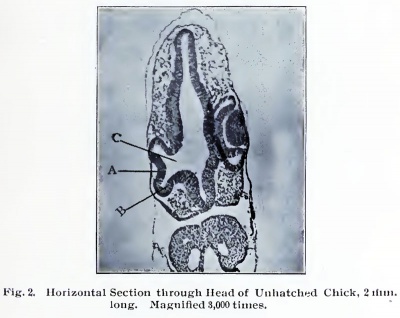
Fig. 2. Horizontal Section through Head of Unhatched Chick, 2 mm long. Magnified 3,000 times.
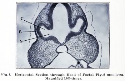
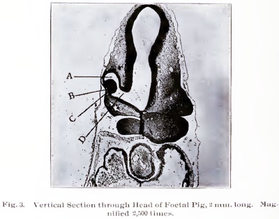
As this stalk grows outward, the anterior portion rises upward, as shown in vertical section at A, Fig. 3. When the optic stalk comes near to the surface, the anterior portion enlarges, as shown at C, Fig. i. Also when the optic stalk encroaches on the surface, it stimulates the epithelial cells forming- the skin and they multiply rapidly (see B, Figs. I and 3), and the anterior wall of the primary optic vesicle invaginates and passes inside of the vesicle. (See A, Fig. 2, and C, Fig. 3.) This invagination might be likened to the denting of a hollow rubber ball.
This invaginated portion forms the secondary optic vesicle and it is from this that the nine innermost layers of the retina are eventually formed, while the primary optic vesicle only forms the outer or pigment layer. As the secondary
Fig. 3. Vertical Section through Head of Foetal Pig, 2 mm long. Magnified 2,500 times.
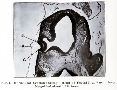
Fig. 4. Horizontal Section through Head of Foetal Pig, 3 mm long. Magnified about 1,200 times. |
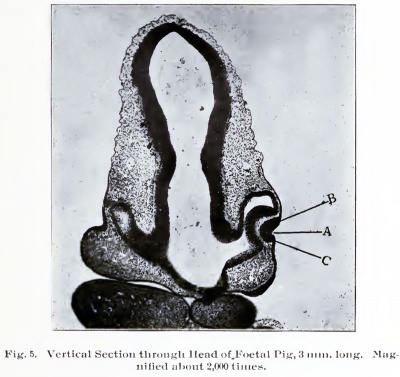
Fig. 5. Vertical Section through Head of Foetal Pig, 3 mm long. Magnified about 2,000 times. |
Optic vesicle is passing into the primary optic vesicle there appears in front of the secondary optic vesicle a depression on the surface at the point of activity of the epithelial cells. (See A, Fig. 4, and A, Fig. 5.) This depression becomes deeper and deeper and the mouth is finally closed by the rapid formation of cells around the depression, as shown at B and C, Figs. 4 and 5.
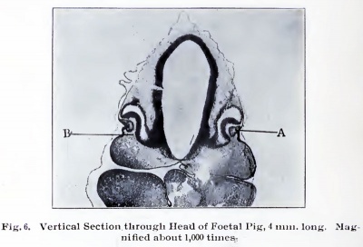
Fig. 6. Vertical Section through Head of Foetal Pig, 4 mm long. Magnified about 1,000 times.
Thus a vesicle is formed which is known as the lens vesicle, as shown at A, Fig. 6. Then this vesicle hecomes separated from the surface and passes into the secondary optic vesicle (see A, Fig. 7), and eventually forms the lens, which will be described later.
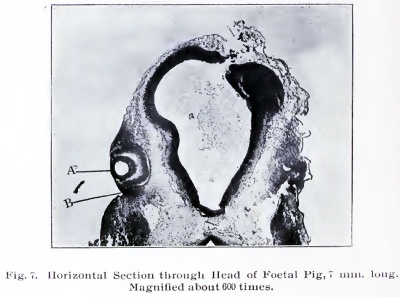
Fig. 7. Horizontal Section through Head of Foetal Pig, 7 mm long. Magnified about 600 times. |
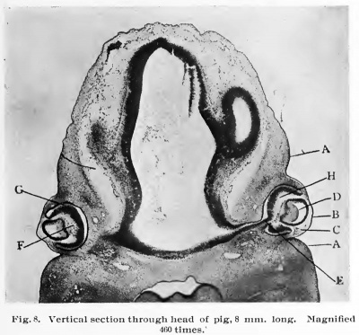
Fig. 8. Vertical section through head of pig, 8 mm. long. Magnified 460 times." |
As the lens vesicle passes into the secondary optic vesicle, some of the mesoblastic cells pass upward from below, behind it (see B, Fig. 6), and it is these mesoblastic cells which will eventually multiply and form the vitreous body. It will be remembered that the mesoblast is the middle layer of the three primary layers, first formed in the foetus. The surface of the skin from which the lens vesicle was cut away, remains and forms the cornea and some students of the embryology of the eye believe that the cornea owes its transparency to the changes that take place in the nature of the epithelial cells during the formation of the lens vesicle from this immediate point. The lids are formed by an external fold, growing downward from above and upward from below the eyeball and the first indication of this growth is shown at B, Fig. 7.
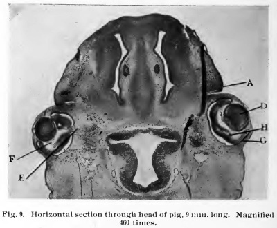
Fig. 9. Horizontal section through head of pig, 9 460 times.
The further development of these folds is shown at A Figs. 8 and 9, also, there is a groove running from the inner side of the eye to the nasal cleft of the foetus and the edges of this groove come together and cover in the cells at the bottom of this groove and a cord is formed from the nasal cleft to the palpebral fissure. (The palpebral fissure is the opening between the lids.) This cord divides and one branch goes to the upper lid and the other to the lower. Later there is a tube formed from this cord and this tube so formed is the lachrymal or tear duct, which runs from the palpebral fissure to the nose, and it is through this duct that the tears are pumped from the conjunctival sack to the nose. This process will be explained later.
In Figs. 7 and 8 the lens vesicle will be seen to have been entirely separated from the surface, and at B, Fig. 8, is seen the epithelial cells which will form the outer layer of the cornea, and at C, Fig. 8, is seen the mesoblastic cells which will multiply and eventually form the four innermost layers of the cornea, the iris and other structures anterior to the lens.
At D, Fig. 8, is seen the commencement of the formation of the lens substance. This formation is accomplished by the cells of the posterior portion of the lens vesicle wall elongating and forming long spindle cells. These grow forward and fill the whole cavity of the lens vesicle, as shown at D, Fig. 9, and these extend from the anterior to the posterior limits of the cavity and are known as the lens fibers. At E, Fig. 8, is seen the opening at the posterior pole of the eye ball, where the axis cylinder processes make their escape from the eye ball, as shown at E, Fig. 9, to pass into the optic nerve as they grow from the retina toward the brain. This opening through which the optic fibers leave the eye ball is known as the choroidal fissure in the adult eye.
At F, Figs. 8 and 9, are shown the mesoblastic cells which have passed into the space between the retina and the lens. As shown at B, Fig. 6, they are just commencing to form the vitreous body, and this cavity so filled is known as the vitreous cavity in the adult eye. At G, Figs. 8 and 9, the primary optic vesicle is shown, which has become quite thin, and in the cells forming it there is being deposited pigment granules, and it will be remembered that this eventually forms the outer or pigment layer of the retina. At H, Figs. 8 and 9, will be seen the first indication of the formation of the nine innermost layers of the retina, and these nine layers are all formed from the walls of the secondary optic vesicle.
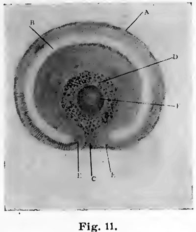
Fig. 11. Colobomba of the fundus in the adult and means a lack of development.
D, Fig. 11, is the vitreous cavity. F, Fig. 11, is the lens vesicle cut through, and the two edges which come together and close the choroidal fissure are shown at E. When this union fails to take place we have an anomaly known as
|
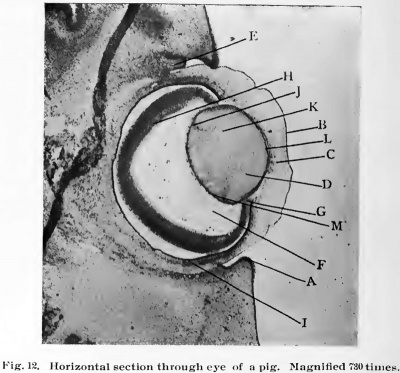
Fig. 12. Horizontal section through eye of a pig. Magnified 730 times. |
At A, Fig. 12, will be seen the further development of the lids; B, Fig. 12, the anterior epithehal layer of the cornea, and just beneath it is seen a lighter colored line. This is the anterior homogeneous (structureless) layer of the cornea, also known as Bowman's membrane, as he was the first to describe it. C, Fig. 12, shows the lamina propria (proper layer) of the cornea. D, Figs. 12 and 13, shows the lens fibers extending from the front to the back of the lens. These fibers are simply long spindle cells and each one has a nucleus. These form a crescent-shaped line of dots, as seen at K, Figs. 12 and 13, running from one side of the lens to the other. At J, Figs. 12 and 13, is seen the transitional (transformation) zone, and it is at this point that the lens fibers are formed, and this formation is simply the multiplication of the columnar epithelial cells, which first formed the wall of the lens vescicle and their elaboration into spindle cells. These spindle cells are known as the lens fibers. |
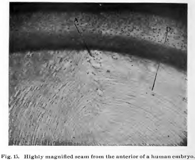
These fibers are especially well illustrated at D, Fig. 15, and anterior to this transitional zone where the lens fibers are formed in the adult eye will be found a single layer of the columnar epithelial cells. Underneath the capsule L, Figs. 12 and 13, and J, Fig. 16, while posterior to the transitional zone, no such cells will be found, they having elongated to form the lens fibers, as shown at D, Figs. 8 to 15.
Fig. 13. Human embryo eye, 2 months. Magnified 1,080 times.
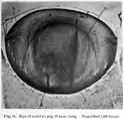
Fig. 14. Eye of embryo pig, 10 mm long. Magnified 1,600 times.
After the lens vesicle is completely filled by fibers, extending from the front to the back of the lens in an anteriorposterior direction, as shown by the lens in Figs. 9 to 13, there is a continuation of growth of the lens by the multiplication and elongation of the columnar cells at the transitional zone, J, Figs. 12, 13 and 14. These grow forward and backward toward the anterior and posterior poles of the lens, around the ends of the first formed fibers, and these latterly developed fibers form the soft outer or cortical portion of the lens, B, Fig. 15. While the first formed fibers constitute the nuclear or central denser portion of the lens, C, Fig. 14, these last formed fibers grow in such a way that when their ends come into apposition at the front and back of the lens, there are formed seams, as shown at A, Figs. 14 and 15. A, Fig. 14, is at the posterior surface of an embryo pig, and A, Fig. 15, is a more highly magnified seam from the anterior of a human embryo. These seams have a star or stilate shape, the central part being at the anterior and posterior poles and the points run outward toward the equator of the lens, and it is these seams, where the ends of the fibers in the cortical portion of the lens abut against each other, that forms the so-called lens stars. These fibers of the lens are long, diamond-shaped, spindle cells, and these are arranged in lamella or layers and all held together by a matrix of gellatinous cement substance, and when a lens is macerated (soaked) in an alkaline solution, which will dissolve this cement substance, these lamella of the lens may be peeled ofif, and the best illustration is the peeling of the layers of an onion. At E, Fig. 12, is seen the commencement of the growth of the third eye lid, known as the membrana nictatans (winking membrane) in the lower animals, especially birds. This develops in man up to a certain stage, then ceases and remains as a vestige in a crescent-shaped fold near the inner side of the eye, and is called the plica semilunaris (half moon fold). At F, Fig. 12, is seen the developing vitreous body and the dark spots are the small blood vessels which furnish this body its nutrition during development. These are from the hyaloid artery, which will be described later, and these atrophy before birth. At G, Fig. 12, it will be seen that the brim or fornix at the anterior margin of the primary and secondary optic vesicles are in apposition to the capsule of the lens at its equator, and this enables some of the connective tissue, which binds the retina together, which is known as the fibers of Mueller (he being the first to discover them) to become attached to the capsule of the lens, and as the eye enlarges and the retina settles farther backward, these attached fibers elongate and thus the suspensory ligament (also known as the zonule of Zinn) is formed, and this accounts for this connection between the retina and the lens.
Fig. 15. Highly uiaguitied detail from the anterior of a human embryo,
At H, Fig. 12, will be seen the farther development of the layers of the retina. At I, Fig. 12, are seen some cells, which are showing signs of activity. This is the first sign of the development of the choroid and sclerotic coats.
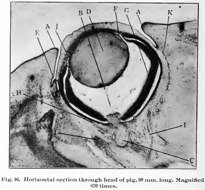
At A, Fig. 16, it will be seen that the lids are gradually covering the cornea and the membrana nictatans. E, Fig. 16, is not any farther developed than seen in Fig. 12 at E.
At B, Fig. 16, will be seen a portion of the hyaloid artery. This is an artery given off by the arteria centralis retina at the head of the optic nerve and only exists during foetal life lor it atrophies before birth. It supplies the nutrition necessary for the development of the vitreous body and the lens and when these are fully matured it atrophies and the canal through which it passed remains as a lymph channel and is known as the hyaloid canal, or the canal of Stilling, in the adult eye. The hyaloid artery, as before stated, is a branch of the arteria centralis retina and runs from the head of the optic nerve to the posterior of the lens, giving off small twigs to the developing vitreous body. At the posterior surface of the lens it breaks up into several branches. These pass around the lens to the front and there come together, forming anastomoses (an anastomosis is where one vessel runs into another and continues by continuity of tissue), and the connective tissue which these vessels are imbedded in forms the pupilary membrane, which will be described later. There is a connection of these hyaloid arteries by anastomosing vessels from the front of the iris, near its free margin, with the branches of the blood vessels of the iris. This connection only exists during foetal life.
Fig. 16. Horiaontal section through head of pig, 20 mm long. Magnified 670 times.
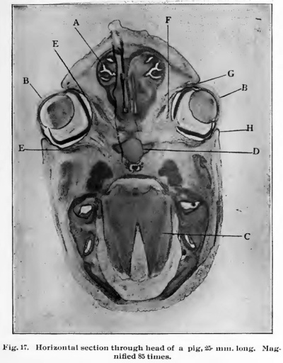
Fig 17. Horizontal section through head of a pig, 25 mm long. Magnified 85 times.
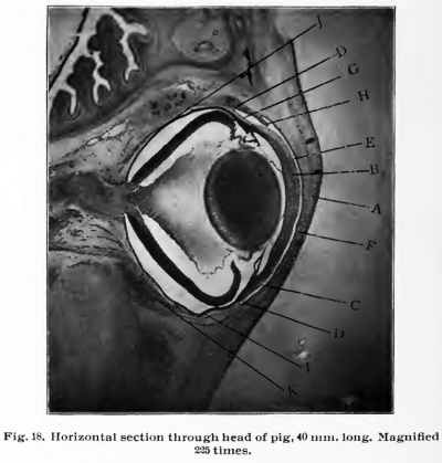
Fig. 18. Horizontal section through head of pig, 40 mm. long. Magnified 225 times.
At F, Fig. 16, is shown a band of tissue connecting the lens with the retina. This will eventually form the suspensory ligament or the Zonule of Zinn of the older writers. At H, Fig. 16, is shown the farther development of the cells which will eventually form the choroid and sclerotic and it will be noted that they may be traced well into the optic nerve (C, Fig. 16), at either side at the choroidal fissure, and it is these cells which will form the lamina cribrosa (seive layer), which strengthens the eye ball at this point in the adult eye. Also C, Fig. 16, shows the first growth of the axis cylinder processes through the choroidal fissure to form the optic nerve.
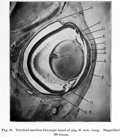
Fig. 19. Vertical section through head of pig, 40 mm long. Magnified 320 times.
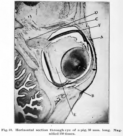
Fig. 20. Horizontal section through eye of a pig, 50 mm long. Magnified 150 times.
It must be remembered that the fibers which transmit impulses of the sight from the retina to the brain, grow from the cells in the ganglionic layer of the retina, back toward the brain and not from the brain to the retina. At I, Fig. 16, is shown the activity of the cells, which are just commencing to form the recti (straight) or extrinsic muscles of the eye. At K, Fig. 16, will be seen a line of small openings. This is the commencement of the space of Tenon. Fig. 17 is a horizontal section through the head of a pig, 25 M. M. long, and is shown to illustrate the rapid development of the eyes in the growth from 9 to 25 M. M. in length. A, Fig. 17, shows the nasal cavities. C, Fig. 17, shows the developing brain, and D shows the developing bone. At E is seen the farther development of the extrinsic muscles, at F is shown the farther development of the choroid and sclerotic and at G is seen the plica semilunaris and at H the lids.
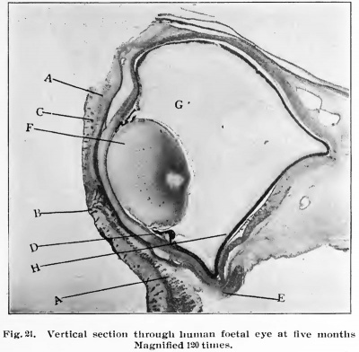
Fig. 21. Vertical section through human foetal eye at five months. Magnified 120 times.
At A, Figs. 18 and 19, are shown the lids entirely covering the front of the eye ball and just back of it the cornea B, and the space between the two, C, is the conjunctival sack.
The conjunctiva lines the inner surface of the lids, then folds on itself as shown at D. This is known as the Fornix (arch) conjunctiva. Then it covers all the front exposed portion of the eye ball except the cornea. The epithelial layer of the conjunctiva, continues over the cornea and forms the outermost or stratified epithelial layer of this structure, E, Figs. i8 and 19. That portion of the conjunctiva lining the lids is known as the palpebral conjunctiva, F. Figs. 18 and 19, and that portion covering the exposed scleral portion of the eye ball is known as the ocular conjunctiva, as shown at G. At H, Fig. 18, is shown the plica semilunaris, and it will be noted that it is gradually becoming smaller.
At I, Figs. 18 and 19, is shown the choroid, which is just forming; at J, is shown the sclerotic and at K, Figs. 18 and 19, is shown the farther development of the extrinsic muscles.
When the two lids come into apposition in front of the eye ball they become cemented together, as shown at h. Fig. 19. In all animals in which the retina is completely developed before birth the lids are separated at birth, but in those animals whose retina is not fully developed at birth, such as the kitten and puppy, the lids do not separate for some days after birth, or until the retina is sufficiently developed so as to withstand the effects of light.
At A, Fig. 20, is shown the conjunctival sack, at B the shrinking plica semilunaris and at C the tendon of the external rectus muscle and its attachment to the eye ball in front and the belly of the muscle posteriorly. At D, Fig. 20, is shown the sheath of the optic nerve and the farther development of the nerve itself, at E the vitreous body and at F it will be seen that the retina is farther developed and about four layers may be made out.
At A, Fig. 21, is shown the developing fibers of the orbicularis (circular) palpebrarum muscle, at B is shown the margins of the lids and the developing cilia (hairs) or eye lashes, and at C is shown a developing hair in the lid. D, Fig. 21, shows the commencement jf the development of the ciliary body and the iris. These are the last structures to be developed within the eye ball. E, Fig. 21, shows the cut end of the inferior oblique muscle, F shows the lens fully developed, also the fibers. G, Fig. 21, shows the vitreous body and H shows the retina practically as is found in the adult eve.
- Next: Anatomy
The Embryology Anatomy and Histology of the Eye: Embryology | Anatomy | Histology | Figures
Glossary Links
- Glossary: A | B | C | D | E | F | G | H | I | J | K | L | M | N | O | P | Q | R | S | T | U | V | W | X | Y | Z | Numbers | Symbols | Term Link
Cite this page: Hill, M.A. (2024, April 27) Embryology Book - The Embryology Anatomy and Histology of the Eye 1. Retrieved from https://embryology.med.unsw.edu.au/embryology/index.php/Book_-_The_Embryology_Anatomy_and_Histology_of_the_Eye_1
- © Dr Mark Hill 2024, UNSW Embryology ISBN: 978 0 7334 2609 4 - UNSW CRICOS Provider Code No. 00098G
