Book - The Elements of Embryology - Mammalian 1
| Embryology - 27 Apr 2024 |
|---|
| Google Translate - select your language from the list shown below (this will open a new external page) |
|
العربية | català | 中文 | 中國傳統的 | français | Deutsche | עִברִית | हिंदी | bahasa Indonesia | italiano | 日本語 | 한국어 | မြန်မာ | Pilipino | Polskie | português | ਪੰਜਾਬੀ ਦੇ | Română | русский | Español | Swahili | Svensk | ไทย | Türkçe | اردو | ייִדיש | Tiếng Việt These external translations are automated and may not be accurate. (More? About Translations) |
Foster M. Balfour FM. Sedgwick A. and Heape W. The Elements of Embryology (1883) Vol. 2. (2nd ed.). London: Macmillan and Co.
| The Elements of Embryology 1883
1 Chicken : Hen's egg and the beginning of incubation | Whole history of incubation | day 1 of incubation | first half of day 2 | second half of day 2 | day 3 | day 4 | day 5 | day 6-21 | Appendix | Figures as Gallery
|
| Historic Disclaimer - information about historic embryology pages |
|---|
| Pages where the terms "Historic" (textbooks, papers, people, recommendations) appear on this site, and sections within pages where this disclaimer appears, indicate that the content and scientific understanding are specific to the time of publication. This means that while some scientific descriptions are still accurate, the terminology and interpretation of the developmental mechanisms reflect the understanding at the time of original publication and those of the preceding periods, these terms, interpretations and recommendations may not reflect our current scientific understanding. (More? Embryology History | Historic Embryology Papers) |
Volume 2 - The History of the Mammalian Embryo
- General Development of the Embryo
- Embryonic Membranes and Yolk-Sac
- Organs from Epiblast
- Organs from Mesoblast
- Alimentary Canal
- Appendix
- Figures
Volume 1 - The History of the Chick
General Development of the Embryo
THERE is a close agreement in the history of the development of the embryo of the various kinds of Mammals. We may therefore take one, the Rabbit, as a type. There are without doubt considerable variations to be met with in the early development even of species nearly allied to the Rabbit, but at present the true value of these variations is not understood, and they need not concern us here.
The ovarian ovum
Mammals possess two ovaries situated in the body cavity, one on either side of the vertebral column immediately posterior to the kidneys. They are somewhat flattened irregularly oval bodies, a portion of the surface being generally raised into protuberances due to projecting follicles.
In an early stage of development the follicle in the mammalian ovary is similar to that of the fowl, and is formed of flat cells derived from the germinal cells adjoining the ovum. As development proceeds however it becomes remarkably modified. These flat cells surrounding the ovum become columnar and then one or two layers deep. Later they become thicker on one side of the ovum than on the other, and there appears in the thickened mass a cavity which gradually becomes more and more distended and filled with an albuminous fluid.
As the cavity enlarges, the OYum, around which are several layers of cells, forms a prominence projecting into it. The follicle cells are known as the membrana granulosa, and the projection in which the ovum lies as the discus or cumulus proligerus. The whole structure with its tunic is known as the Graafian follicle.
If the ovary of a mature female during the breeding season be examined, certain of the protuberances on its surface maybe seen to be considerably larger than others; they are more transparent than their fellows and their outer covering appears more tense ; these are Graafian follicles containing nearly or quite ripe ova. Upon piercing one of these follicles with a needle-point the ovum contained therein spirts forth together with a not inconsiderable amount of clear fluid.
Egg Membranes
The ovum is surrounded by a radiately striated membrane, the zona radiata, internal to which in the nearly ripe egg a delicate membrane has been shown, by Ed. v. Beneden, to exist. The cells of the discus are supported upon an irregular granular membrane external to the zona radiata. This membrane is more or less distinctly separated from the zona, and the mode of its development renders it probable that it is the remnant of the first formed membrane in the young ovum and is therefore the vitelline membrane.
Maturation and impregnation of the ovum
As the ovum placed in the Graafian follicle approaches maturity the germinal vesicle assumes an excentric position and undergoes a series of changes which have not been fully worked out, but which probably are of the same nature as those which have been observed in other types (p. 17). The result of the changes is the formation of one or more polar bodies, and the nucleus of the mature ovum (female pronucleus).
At certain periods one or more follicles containing a ripe ovum burst1 , and their contents are received by the fimbriated extremity of the Fallopian tube which appears according to Hensen to clasp the ovary at the time. The follicle after the exit of the ovum becomes filled with blood and remains as a conspicuous object on the surface of the ovary for some days. It becomes eventually a corpus luteum. The ovum travels slowly down the Fallopian tube. It is still invested by the zona radiata, and in the rabbit an albuminous envelope is formed around it in its passage downwards. Impregnation takes place in the upper part of the Fallopian tube, and is shortly followed by the segmentation, which is remarkable amongst the Amniota for being complete2.
The entrance of the spermatozoon into the ovum and its subsequent fate have not been observed. Van Beneden describes in the rabbit the formation of the first segmentation nucleus (i.e. the nucleus of the ovum after fertilization) from two nuclei, one peripheral and the other ventral, and deduces from his observations that the peripheral nucleus was derived from the spermatic element.
- 1 So far as is known there is no relation between the bursting of the follicle and the act of coition.
- 2 It is stated by Bischoff that shortly after impregnation, and before the commencement of the segmentation, the ova of the rabbit and guinea-pig are covered with cilia and exhibit the phenomenon of rotation. This has not been noticed by other observers.
Segmentation
The process of segmentation occupies in the rabbit about 72 hours; but the time of this and all other stages of development varies considerably in different animals.
The details of segmentation in the rabbit are differently described by various observers ; but at the close of segmentation the ovum appears undoubtedly to be composed of an outer layer of cubical hyaline cells, almost entirely surrounding an inner mass of highly granular rounded or polygonal cells.
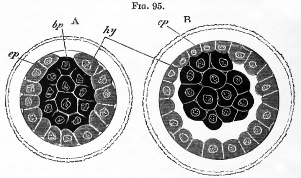
Fig. 95. Optical sections of a rabbit's ovum at two stages closely following upon the segmentation. (After E. van Beneden.)
- ep. outer layer ; hy. inner mass ; bp. Van Beneden's blastopore. The shading of the outer and inner layers is diagrammatic.
In a small circular area however the inner mass of cells remains exposed at the surface (Fig. 95, A). This exposed spot may for convenience be called with v. Beneden the blastopore, though, as will be seen by the account given of the subsequent development, it in no way corresponds with the blastopore of other vertebrate ova.
In the following account of the segmentation of the rabbit's ovum, v. Beneden's description is followed as far as the details are concerned, his nomenclature is however not adhered to1.
According to v. Beneden the ovum first divides into two nearly equal spheres, of which one is slightly larger and more transparent than the other. The larger sphere and its products will be spoken of as the outer spheres, and the smaller one and its products as the inner spheres, in accordance with their different destinations.
Both the spheres are soon divided into two, and each of the four so formed into two again ; and thus a stage with eight spheres ensues. At the moment of their first separation these spheres are spherical, and arranged in two layers, one of them formed of the four outer, and the other of the four inner spheres. This position is not long retained, for one of the inner spheres passes to the centre ; and the whole ovum again takes a spherical form.
In the next phase of segmentation each of the four outer spheres divides into two, and the ovum thus becomes constituted of twelve spheres, eight outer and four inner. The outer spheres have now become markedly smaller than the inner.
The four inner spheres next divide giving rise, together with the eight outer spheres, to sixteen spheres in all ; which are nearly uniform in size. Of the eight inner spheres four soon pass to the centre, while the eight now superficial outer spheres form a kind of cup partially enclosing the inner spheres. The outer spheres now divide in their turn, giving rise to sixteen spheres which largely enclose the inner spheres. The segmentation of both outer and inner spheres continues, and in the course of it the outer spheres spread further and further over the inner, so that at the close of segmentation the inner spheres constitute a central solid mass almost entirely surrounded by the outer spheres. In a small circular area however the inner mass of spheres remain for some time exposed at the surface (Fig. 95 A).
- 1 The cells spoken of as the outer layer correspond to Van Beneden's epiblast, whilst those cells spoken of as the inner correspond to his primitive hypoblast.
The blastodemic vesicle
After its segmentation the ovum passes into the uterus. The outer cells soon grow over the blastopore and thus form a complete superficial layer. A series of changes next take place which result in the formation of what has been called the blastodermic vesicle.
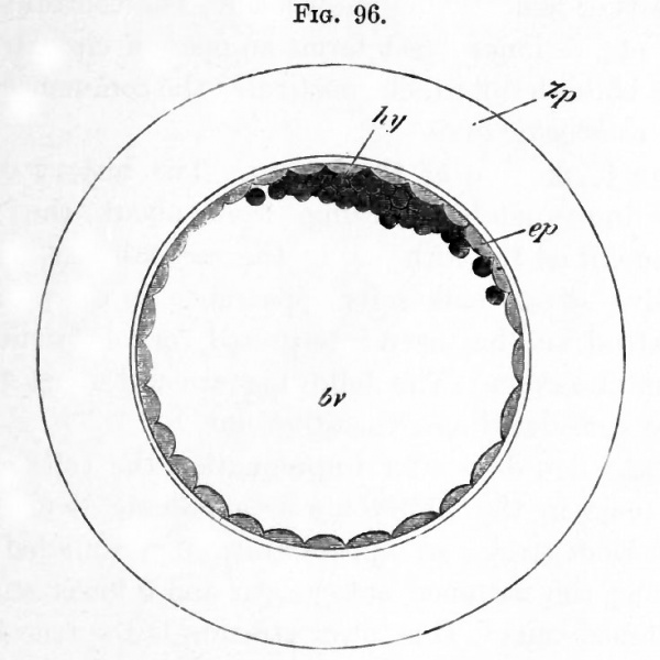
These changes commence with the appearance of a narrow cavity between the outer and inner layers, which extends so as completely to separate them except in the region adjoining the original site of the blastopore (Fig. 95 B)1. The cavity so formed rapidly enlarges, and with it the ovum also ; so that this soon takes the form of a thin walled vesicle with a large central cavity. This vesicle is the blastodermic vesicle. The greater part of its walls are formed of a single row of flattened outer layer cells; while the inner mass of cells forms a small lens-shaped mass attached to the inner side of the outer layer (Fig. 96).
Although by this stage, which occurs in the rabbit between seventy and ninety hours after impregnation, the blastodermic vesicle has by no means attained its greatest dimensions, it has nevertheless grown from about 0.09 mm. the size of the ovum at the close segmentation to about 0.28 in diameter. It is enclosed by the zona radiata and the albuminous layer around it. The blastodermic vesicle continues to enlarge rapidly, and during the process the inner mass undergoes important changes. It spreads out on the inner side of the outer layer and at the same time loses its lens-like form and becomes flattened. The central part of it remains however thicker, and is constituted of two rows of cells, while the peripheral part, the outer boundary of which is irregular, is formed of an imperfect layer of amoeboid cells which continually spread further and further beneath the outer layer. The central thickening of the inner layer forms an opaque circular spot on the blastoderm, which constitutes the commencement of the embryonic area.
- 1Van Beheden regards it as probable that the blastopore is situated somewhat excentrically in relation to the area of attachment of the inner mass to the outer layer.
Fig. 96 Rabbit's ovum between 70 90 hours after impregnation. (After E. van Beneden.)
- bv. cavity of blastodermic vesicle (yolk-sac) ; ep. outer layer ; hy. inner mass ; Zp. albuminous envelope.
The formation of the layers
The history of the stages immediately following, from about the commencement of the fifth day to the seventh day, when a primitive streak makes its appearance, is not perfectly understood, and has been interpreted very differently by various observers. The following account must therefore be considered as a tentative one.
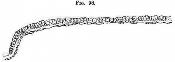
About five days after impregnation the cells of the inner mass in the embryonic area become divided into two distinct strata, an upper stratum of rounded cells adjoining the flattened outer layer and a lower stratum of flattened cells. This lower stratum is the true hypoblast (Fig. 97). At the edge of the embryonic area the hypoblast is continuous with a peripheral ring of the amosboid cells of the earlier stage, which now form, except at the edge of the ring, a continuous layer of flattened cells in contact with the outer layer. During the sixth day the middle layer becomes fused with the outer layer, and gives rise to a layer of cells which are columnar and are arranged in the rabbit in a single row (Fig. 98). They form together the true epiblast of the embryonic area.
At this stage therefore the embryonic area, which is circular, is formed throughout of two single layers of cells, a columnar epiblast and a layer of flattened hypoblast.
Fig. 97. Section through the nearly circular embryonic area of a rabbit ovum of six days. (From Allen Thomson, after E. van Beneden.)
- ect. upper layer ; mes. middle layer ; ent. true hypoblast.
Fig. 98. Section through the blastoderm of a rabbitt on the seventh day: taken in front of the primitive streak.
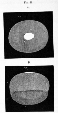
- Half of the area is represented. Towards the end of the sixth day the embryonic area of the rabbit, which has hitherto been round, becomes oval. A diagrammatic view of the whole blastodermic vesicle at about the beginning of the seventh day is given in Fig. 99. The embryonic area is represented in white. The line ge in B shows the extension of the hypoblast round the inside of the vesicle.
Fig. 99. Views of the Blastodermic Veiscle of a Rabbit on the Seventh Day without the Zona.
- A. from above, B. from the side. (From Kolliker.) ag. embryonic area ; ge. boundary of the hypoblast.
The biastodermic vesicle is therefore formed of three areas, (1) the embryonic area with two layers, a columnar epiblast and flat hypoblast; (2) the region around the embryonic area where the walls of the vesicle are formed of flattened epiblast 1 and of hypoblast ; (3) the area beyond this again where the vesicle is formed of flattened epiblast 1 only.
The changes which next take place begin with the formation of a primitive streak, homologous with, and in most respects similar to, the primitive streak in Birds.
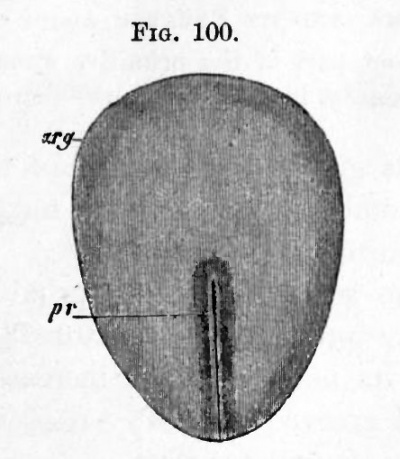
Fig. 100. Embryonic Area of an Eight Days' Rabbit.
- (After Kolliker.) arg. embryonic area ; pr. primitive streak.
The formation of the streak is preceded by that of a dark spot near the middle of the blastoderm, forming the nodal point of Hensen. This spot subsequently constitutes the front end of the primitive streak.
Early on the seventh day the embryonic area becomes pyriform, and at its posterior and narrower end the primitive streak makes its appearance ; it is due to a proliferation of rounded cells from the epiblast.
(1The epiblast of the blastodermic vesicle beyond the embryonic area is formed of the outer layer only.)
Fig. 101. Section through an Oval Blastoderm or a Rabbit on the Seventh Day.
- The length of the area was about 1.2 mm. and its breadth about .86 mm. Through the front part of the primitive streak ; ep. epiblast ; m. mesoblast ; hy. hypoblast ; pr. primitive streak.
These cells give rise to a part of the mesoblastic layer of the embryo, and may be termed from their origin the primitive streak mesoblast.
During the seventh day the primitive streak becomes a more pronounced structure (Fig. 101), the mesoblast in its neighbourhood increases in quantity, while an axial groove (Fig. 100) the primitive groove is formed on its upper surface.
The formation of the medullary groove
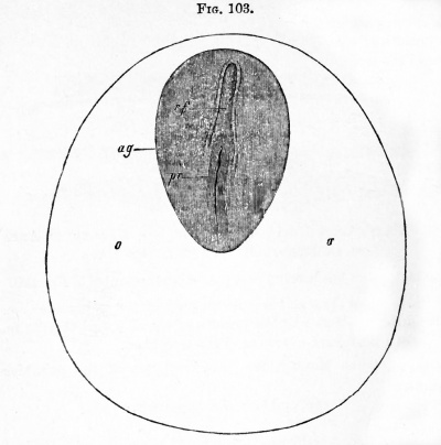
In the part of the embryonic area in front of the primitive streak there arise during the eighth day two folds bounding a shallow median groove, which meet in front, but diverge behind, and enclose between them the foremost end of the primitive streak (Fig. 103). These folds are the medullary folds and they constitute the first definite traces of the embryo. The medullary plate bounded by them rapidly grows in length, the primitive streak always remaining at its hinder end.
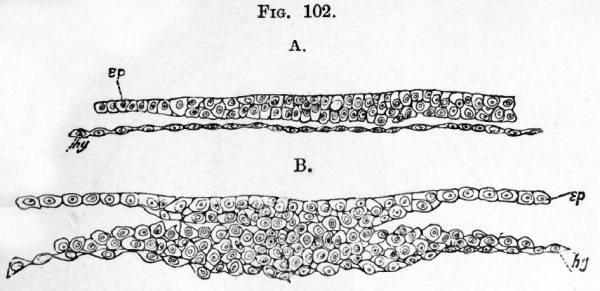
FIG. 102. Two transverse suctions through the embryonic area of an embryo rabbit of seven day. The embryo has nearly the appearance represented in Fig. 100. ep. epiblast ; hy. hypoblast.
- A. is taken through the anterior part of the embryonic area. It represents about half the breadth of the area, and there is no trace of a medullary groove or of the mesoblast.
- B. is taken through the posterior part of the primitive streak.
While the lateral epiblast is formed of several rows of cells, that of the medullary plate is at first formed of but a single row (Fig. 104, mg).
The mesoblast and notochord
The mesoblast in mammalia has, as in the chick, a double origin, and the details of its development appear to resemble essentially those in the chick. It arises (1) from the epiblast of the primitive streak ; this has been already described ; (2) from the primitive hypoblast in front and at the sides of the primitive streak. The latter is known as hypoblastic mesoblast, and as in the chick appears to originate as two lateral plates split off from the primitive hypoblast.
Fig. 103. Embryonic area of a seven days' embryo rabbit. (From Kolliker.) o. place of future area vasculosa ; rf. medullary groove ; pr. primitive streak ; ag. embryonic area. In the region o. a layer of mesoblast has already grown ; there are however as yet no signs of blood-vessels in it. This mesoblast is derived from the mesoblast of the primitive streak (Kolliker).
These two plates are at first continuous in the axial line with the primitive hypoblast. When the medullary groove is formed the lateral bands of mesoblast become separate from the axial hypoblast and give rise to two independent lateral plates of mesoblast(Fig. 104). The axial band of hypoblast eventually oives rise to the notochord.
Fig. 104. Transverse section through an embryo rabbit of eight days. epi. epiblast ; me. mesoblast ; hy. hypoblast ; mg. medullary groove.
The mesoblastic elements from these two sources, though at first characterised by the difference in the appearance of their cells (Fig. 102, B), those of the primitive streak mesoblast being more rounded, soon become blended and indistinguishable from one another; so that it is difficult to say to what parts of the fully formed mesoblast they severally contribute.
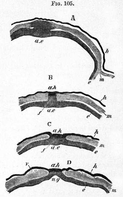
In tracing the changes which take place in the relations of the layers, while passing from the region of the embryo to that of the primitive streak, it will be convenient to follow the account given by Schafer for the guinea-pig, which on this point is far fuller and more satisfactory than that of other observers. In doing so we shall leave out of consideration the fact that the layers in the guinea-pig are inverted. Fig. 105 represents a series of sections through this part in the guineapig. The anterior section (D) passes through the medullary groove near its hinder end. The commencement of the primitive streak is marked by a slight prominence on the floor of the medullary groove between the two diverging medullary folds (Fig. 105 C, ae).
Fig. 105. A series of transverse sections through the junction of the primitive streak and medullary groove of a young guinea-pig. (After Schafer.) A. is the posterior section. e. epiblast ; m. mesoblast ; h. hypoblast ; ae. axial epiblast of the primitive streak ; ah. axial hypoblast attached in B. and C. to the epiblast at the rudimentary blastopore ; mg. medullary groove ; /. rudimentary blastopore.
Where this prominence becomes first apparent the epiblast and hypoblast are united together. The mesoblast plates at the two sides remain in the meantime quite free. Slightly further back, but before the primitive groove is reached, the epiblast and hypoblast are connected together by a cord of cells (Fig. 105 B), which in the section next following becomes detached from the hypoblast and forms a solid keel projecting from the epiblast. In the following section the hitherto independent mesoblast plates become united with this keel (Fig. 105 A) ; and in the posterior sections, through the part of the primitive streak with the primitive groove, the epiblast and mesoblast continue to be united in the axial line, but the hypoblast remains distinct. These peculiar relations may shortly be described by saying that in the axial line the hypoblast becomes united with the epiblast at the posterior end of the embryo; and that the cells which connect the hypoblast and epiblast are posteriorly continuous with the fused epiblast and mesoblast of the primitive streak, the hypoblast in the region of the primitive streak having become distinct from the other layers.
The notochord
The thickened axial portion of the hypoblast in the region of the embryo becomes separated, as we have already pointed out, from the lateral parts as the notochord.
Very shortly after the formation of the notochord, the hypoblast grows in from the two sides, and becomes quite continuous across the middle line. The formation of the notochord takes place from before backwards; and at the hinder end of the embryo it is continued into the mass of cells which forms the axis of the primitive streak, becoming therefore at this point continuous with the epiblast. The notochord in fact behaves exactly as did the axial hypoblast in the earlier stage.
The peculiar relations just mentioned are precisely similar to those we have already described in the chick (p. 60). They receive their explanation by comparison with the lower types.
The cells which form the junction between the epiblast and the axial hypoblast constitute in the lower types the front wall of a passage perforating the blastoderm and leading from the exterior into the alimentary canal. This passage is the vertebrate blastopore.
In the chick we have seen (p. 72) this passage is present at a certain stage of development as the neurenteric canal ; and in the duck at a still earlier stage. It is also present at an early stage in the mole.
The presence of this blastopore renders it clear that the blastopore discovered by Ed. van Beneden cannot have the meaning he assigned to it in comparing it with the blastopore of the
To recapitulate. At the stage we have now reached the three layers are definitely established.
The epiblast is derived partly from the outer layer of segmentation spheres and partly from the larger proportion of those segmentation spheres which constitute the inner mass. The hypoblast arises from the few remaining cells of the inner mass ; while the mesoblast has its origin partially from the epiblast of the primitive streak and partially from the hypoblast cells anterior to the primitive streak.
During the period in which these changes have been taking place, the rudiments of a vascular area become formed, and while as Kolliker has shewn, the mesoblast of this portion is to some extent derived from the mesoblast of the primitive streak, it is possible that a portion of it owes its origin to hypoblastic mesoblast.
General growth of the embryo
We have seen that the blastodermic vesicle becomes divided at an early stage of development into an embryonic area, and a non-embryonic portion. The embryonic area gives rise to the whole of the body of the embryo, while the non-embryonic part forms an appendage known as the umbilical vesicle, which becomes gradually folded off from the embryo, and has precisely the relations of the yolk-sac of the chick. It is almost certain that the Mammalia are descended from ancestors, the embryos of which had large yolk-sacs, but that the yolk has become reduced in quantity owing to the nutriment received from the wall of the uterus taking the place of that originally supplied by the yolk. A rudiment of the yolk-sac being thus retained in the umbilical vesicle, this structure may be called indifferently umbilical vesicle or yolk-sac.
The yolk which fills the yolk-sac in Birds is replaced in Mammals by a coagulable fluid; while the gradual extension of the hypoblast round the wall of the blastodermic vesicle, which has already been described, is of the same nature as the growth of the hypoblast round the yolk-sac in Birds.
The whole embryonic area would seem to be employed in the formation of the body of the embryo. Its long axis has no very definite relation to that of the blastodermic vesicle. The first external trace of the embryo to appear is the medullary plate, bounded by the medullary folds, and occupying at first the anterior half of the -embryonic area (Fig. 103). The two medullary folds diverge behind and enclose the front end of the primitive streak. As the embryo elongates the medullary folds nearly meet behind and so cut off the front portion of the primitive streak, which then appears as a projection in the hind end of the medullary groove. At the hind end of the medullary groove (mole) a deep pit perforates its floor and enters the mass of mesoblast cells lying below. The pit is a rudiment of the blastopore (described on p. 326) which has been enclosed by the medullary folds.
Henceforward the general course of development is very similar to that in the chick and so will be only briefly described. The special features in the development of particular organs will be described later. In an embryo rabbit, eight days after impregnation, the medullary groove is about 1.80 mm. in length. At this stage a division may be clearly seen in the lateral plates of mesoblast into a vertebral zone adjoining the embryo and a more peripheral lateral zone ; and in the vertebra] zone indications of two somites, about 0.37 mm. from the hinder end of the embryo, become apparent. The foremost of these somites marks the junction, or very nearly so, of the cephalic region and trunk. The small size of the latter as compared with the former is very striking, but is characteristic of Vertebrates generally. The trunk gradually elongates relatively to the head, by the addition behind of fresh somites. The embryo has not yet begun to be folded off from the yolk-sac.
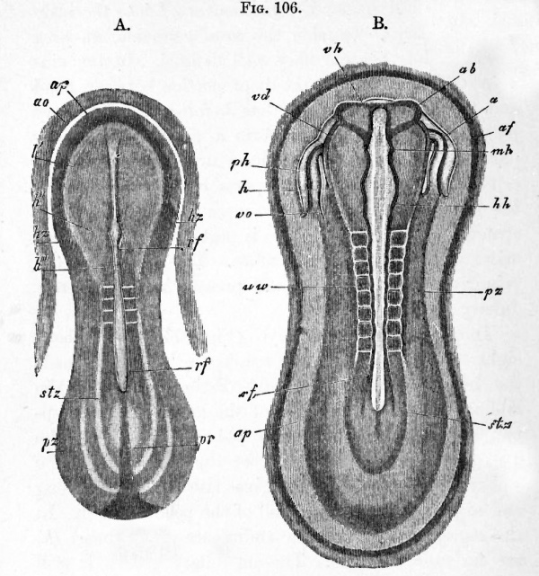
In a slightly older embryo of nine days there appears (Hensen, Kolliker) round the embryonic area a delicate clear ring which is narrower in front than behind (Fig. 106 A. ap). This ring is regarded by these authors as representing the peripheral part of the area pellucida of Birds, which does not become converted into the body of the embryo. Outside the area pellucida, an area vasculosa has become very well defined. In the embryo itself (Fig. 106 A) the disproportion between head and trunk is less marked than before; the medullary plate dilates anteriorly to form a spatula-shaped cephalic enlargement; and three or four somites are established. In the lateral parts of the mesoblast of the head there may be seen on each side a tube-like structure (hz). Each of these is part of the heart, which arises as two independent tubes. The remains of the primitive streak (pr) are still present behind the medullary groove.
In somewhat older embryos (Fig. 106 B) with about eight somites, in which the trunk considerably exceeds the head in length, the first distinct traces of the folding off of the head end of the embryo become apparent, and somewhat later a fold also appears at the hind end. In the formation of the hind end of the embryo the primitive streak gives rise to a tail swelling and to part of the ventral wall of the post-anal gut. In the region of the head the rudiments of the heart (h) are far more definite. The medullary groove is still open for its whole length, but in the head it exhibits a series of well-marked dilatations. The foremost of these (vh) is the rudiment of the fore-brain from the sides of which there project the two optic vesicles (all) ; the next is the mid-brain (mK) and the last is the hind-brain (hh), which is again divided into smaller lobes by successive constrictions. The medullary groove behind the region of the somites dilates into an embryonic sinus rhomboidalis like that of the bird. Traces of the amnion (of) are now apparent both in front of and behind the embryo.
Fig. 106. Embryo rabbits of about nine days from the dorsal side. (From Kolliker.) A. magnified 22 times, and B. 21 times.
- ap. area pellucida ; rf. medullary groove ; h f . medullary plate in the region of the future fore-brain ; h". medullary plate in the region of the future mid-brain ; vh. fore- brain ; ab. optic vesicle ; mh. mid-brain ; lili. and h'". hind-brain ; uw. mesoblastic somite ; stz. vertebral zone ; pz. lateral zone ; liz. and h. heart ; ph. pericardial section of body-cavity ; vo. vitelline vein ; af. amnion fold.
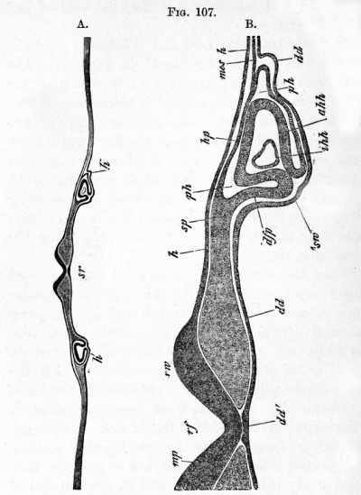
The structure of the head and the formation of the heart at this age are illustrated in Fig. 107. The widely open medullary groove (rf) is shewn in the centre. Below it the hypoblast is thickened to form the notochord dd' ; and at the sides are seen the two tubes, which, on the folding-in of the fore-gut, give rise to the unpaired heart1. Each of these is formed of an outer muscular tube of splanchnic mesoblast (ahh), not quite closed towards the hypoblast, and an inner epithelioid layer (ihh), and is placed in a special section of the body cavity (ph), which afterwards forms the pericardial cavity.
Before the ninth day is completed great external changes are usually effected. The medullary groove becomes closed for its whole length with the exception of a small posterior portion. The closure commences, as in Birds, in the region of the mid-brain. Anteriorly the folding-off of the embryo proceeds so far that the head becomes quite free, and a considerable portion of the throat, ending blindly in front, becomes established. In the course of this folding the, at first widely separated, halves of the heart are brought together, coalesce on the ventral side of the throat, and so give rise to a median undivided heart. The fold at the tail end of the embryo progresses considerably, and during its advance the allantois is formed in the same way as in Birds. The somites increase in number to about twelve. The amniotic folds nearly meet above the embryo.
(1 The details of the development of the heart are described below (ch. xii.).
Fig. 107. Transverse section through the head of a rabbit of the same age as fig. 106 B. (From Kolliker.) B. is a more highly magnified representation of part of A.
- rf. medullary groove ; mp. medullary plate ; rw. medullary fold ; h. epiblast ; dd. hypoblast ; dd. notochordal thickening of hypoblast ; sp. undivided mesoblast ; hp. somatic mesoblast ; sp. splanchnic mesoblast; ph. pericardial section of bodycavity ; ahh. muscular wall of heart ; ihh. epithelioid layer of heart ; mes. lateral undivided mesoblast ; sw. fold of hypoblast which will form the ventral wall of the pharynx ; sr. commencing throat.
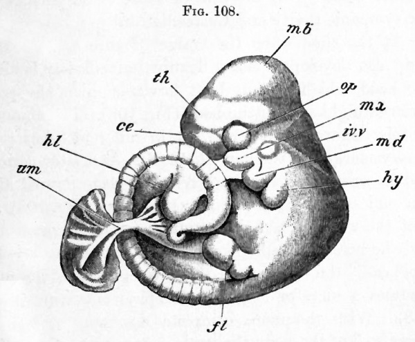
The later stages in the development proceed in the main in the same manner as in the Bird. The cranial flexure soon becomes very marked, the mid-brain forming the end of the long axis of the embryo (Fig. 108). The sense organs have the usual development. Under the fore -brain appears an epiblastic involution giving rise both to the mouth and to the pituitary body. Behind the mouth are three well marked pairs of visceral arches. The first of these is the mandibular arch (Fig. 108 md) y which meets its fellow in the middle line, and forms the posterior boundary of the mouth. It sends forward on each side a superior maxillary process (mx) which partially forms the anterior margin of the mouth. Behind the mandibular arch are present a well-developed hyoid (hy) and a first branchial arch (not shewn in Fig. 108). There are four clefts, as in the chick, but the fourth is not bounded behind by a definite arch. Only the first of these clefts persists as the tympanic cavity and Eustachian tube.
Fig. 108. Advanced embryo of a rabbit (about twelve days). This figure was drawn by Mr Weldon.
- mb. mid-brain ; ih. thalamencephalon ; ce. cerebral hemisphere ; op. eye ; iv.v. fourth ventricle ; moc. maxillary process ; md. mandibular arch ; Jiy. hyoid arch ; fl. fore-limb ; hi. hindlimb ; urn. umbilical stalk.
At the time when the cranial flexure appears, the body also develops a sharp flexure immediately behind the head, which is thus bent forwards upon the posterior straight part of the body (Fig. 108). The amount of this flexure varies somewhat in different forms. It is very marked in the dog (Bischoff ). At a later period, and in some species even before the stage figured, the tail end of the body also becomes bent (Fig. 108), so that the whole dorsal side assumes a convex curvature, and the head and tail become closely approximated. In most cases the embryo, on the development of the tail, assumes a more or less definite spiral curvature (Fig. 108). With the more complete development of the lower wall of the body the ventral flexure partially disappears, but remains more or less persistent till near the close of intra-uterine life. The limbs are formed as simple buds in the same manner as in Birds. The buds of the hind-limbs are directed somewhat forwards, and those of the fore-limb backwards.
The human embryo
Our knowledge as to the early development of the human embryo is in an unsatisfactory state. The positive facts we know are comparatively few, and it is not possible to construct from them a history of the development which is capable of satisfactory comparison with that in other forms, unless all the early embryos known are to be regarded as abnormal. The most remarkable feature in the development, which was first clearly brought to light by Allen Thomson in 1839, is the very early appearance of branched villi. In the last few years several ova, even younger than those described by Allen Thomson, have been met with, which exhibit this peculiarity.
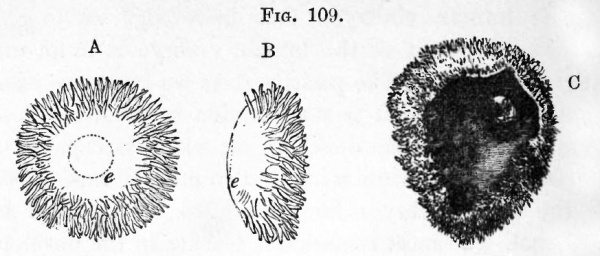
The best preserved of these ova is one described by Reichert1. This ovum, though probably not more than thirteen days old, was completely enclosed by a decidua reflexa. It had (Fig. 109 A and B) a flattened oval form, measuring in its two diameters 5 '5 mm. and 3*5 mm. The edge was covered with branched villi, while in the centre of each of the flattened surfaces there was a spot free from villi. On the surface adjoining the uterine wall was a darker area (e) formed of two layers of cells. Nothing certain has been made out about the structure of ova of this age.
The villi, which at first leave the flattened poles free, seem soon to extend first over one of the flat sides and finally over the whole ovum (Fig. 109 C).
(1Abhandlungen der Konigl. Akad. d. Wiss. zu Berlin, 1873.)
Fig. 109. The human ova during early stages of development. (From Quain's Anatomy.)
- A. and B. Front and side view of an ovum figured by Keichert, supposed to be about thirteen days. e. embryonic area. C. An ovum of about four or five weeks shewing the general structure of the ovum before the formation of the placenta. Part of the wall of the ovum is removed to shew the embryo in situ. (After Allen Thomson.)
Unless the two-layered region of Reichert's ovum is the embryonic area, nothing which can clearly be identified as an embryo has been detected in these early ova. In an ovum described by Breus, and in one described long ago by Wharton-Jones, a mass found in the interior of the ovum may perhaps be interpreted (His) as the remains of the yolk. It is, however, very probable that all the early ova so far obtained are more or less pathological.
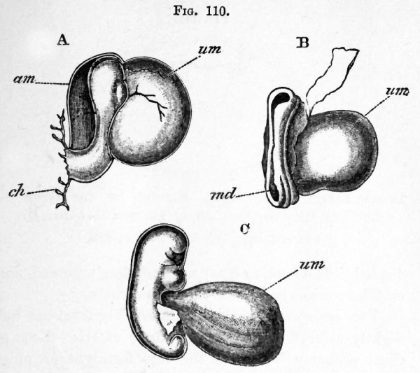
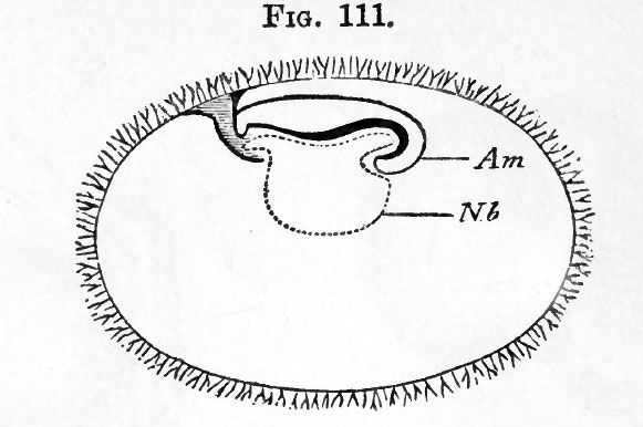
The youngest ovum with a distinct embryo is one described by His. This ovum, which is diagrammatically represented in Fig. Ill in longitudinal section, had the form of an oval vesicle completely covered by villi, being about 8.5 mm. and 5.5 mm. in its two diameters, and flatter on one side than on the other. An embryo with a yolk-sac was attached to the inner side of the flatter wall of the vesicle by a stalk, which must be regarded as the allantoic stalk; the embryo and yolk-sac filled up but a very small part of the whole cavity of the vesicle.
Fig. 110. Three early human embryos. (Copied from His.)
- A. Side view of an early embryo described by His.
- B. Embryo of about 12 14 days described by Allen Thomson.
- C. Young embryo described by His.
- am. amnion ; md. medullary groove ; um. umbilical vesicle ; ck. chorion, to which the embryo is attached by a stalk.
The embryo, which was probably not quite normal (Fig. 110 A), was very imperfectly developed; a medullary plate was hardly indicated, and, though the mesoblast was unsegmented, the head fold, separating the embryo from the yolk-sac (um), was already indicated. The amnion (am) was completely formed, and vitelline vessels had made their appearance.
Fig. 111. Diagrammatic longitudinal section of the ovum to which the embryo (Fig. 110 a.) belonged. (After His.) am. amnion ; Nb. umbilical vesicle.
Two embryos described by Allen Thomson are but slightly older than the above embryo of His. Both of them probably belong to the first fortnight of pregnancy. In both cases the embryo was more or less folded off from the yolk-sac, and in one of them the medullary groove was still widely open, except in the region of the neck (Fig. 110 B). The allantoic stalk, if present, was not clearly made out, and the condition of the amnion was also not fully studied. The smaller of the two ova was just 6 mm. in its largest diameter, and was nearly completely covered with simple villi, more developed on one side than on the other.
In a somewhat later period, about the stage of a chick at the end of the second day, the medullary folds are completely closed, the region of the brain already marked, and the cranial flexure commencing. The mesoblast is divided up into numerous somites, and the mandibular and first two branchial arches are indicated.
The embryo is still but incompletely folded off from the yolk-sac below.
In a still older stage the cranial flexure becomes still more pronounced, placing the mid-brain at the end of the long axis of the body. The body also begins to be ventrally curved (Fig. 110 C).
Externally human embryos at this age are characterized by the small size of the anterior end of the head.
The flexure goes on gradually increasing, and in the third week of pregnancy in embryos of about 4 mm. the limbs make their appearance.
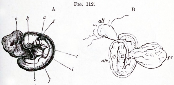
The embryo at this stage (Fig. 112), which is about equivalent to that of a chick on the fourth day, resembles in almost every respect the normal embryos of the Amniota. The cranial flexure is as pronounced as usual, and the cerebral region has now fully the normal size. The whole body soon becomes flexed ventrally, and also somewhat spirally. The yolk-sac (B ; ys) forms a small spherical appendage with a long wide stalk, and the embryo is attached by an allantoic stalk with a slight swelling, probably indicating the presence of a small hypoblastic diverticulum, to the inner face of the chorion.
Fig. 112. Two views of a human embryo of between the third and fourth week.
- A. Side view. (From Kolliker ; after Allen Thomson.) a. amnion ; b. umbilical vesicle ; c. mandibular arch ; e. hyoid arch; /. commencing anterior limb; g. primitive auditory vesicle ; h. eye ; i. heart.
- B. Dorsal view to shew the attachment of the dilated allantoic stalk to the chorion. (From a sketch by Allen Thomson.) am. amnion ; all. allantois ; ys. yolk-sac.
A detailed history of the further development of the human embryo does not fall within the province of this work; while the later changes in the embryonic membranes will be dealt with in the next chapter. For the changes which take place on the formation of the face we may refer the reader to Fig. 113. For a full discussion as to the relation between the human embryos just described and those of other Mammals, we refer the reader to the Comp. Embryology, Vol. II. p. 224 et seq. The guinea pig, rat and mouse present a peculiar method of development, the details of which are not entirely understood, and we do not propose to examine them here. Suffice it to say that the mode of development gives rise to the so-called inversion of the layers; so called because the outer layer of the embryonic vesicle appeared to the older observers to be formed of hypoblast and the embryonic epiblast to be enclosed within.
Fig. 113. Figures shewing the early changes in the form of the human head. (From Quain's Anatomy.)
- A. Head of an embryo of about four weeks. (After Allen Thomson.)
- B. Head of an embryo of about six weeks. (After Ecker.)
- C. Head of an embryo of about nine weeks.
- 1. mandibular arch ; 1'. persistent part of hyomandibular cleft ; a. auditory vesicle.
The Elements of Embryology - Volume 2 (1883)
The History of the Mammalian Embryo: General Development | Embryonic Membranes and Yolk-Sac | Organs from Epiblast | Organs from Mesoblast | Alimentary Canal | Appendix | Figures
| Historic Disclaimer - information about historic embryology pages |
|---|
| Pages where the terms "Historic" (textbooks, papers, people, recommendations) appear on this site, and sections within pages where this disclaimer appears, indicate that the content and scientific understanding are specific to the time of publication. This means that while some scientific descriptions are still accurate, the terminology and interpretation of the developmental mechanisms reflect the understanding at the time of original publication and those of the preceding periods, these terms, interpretations and recommendations may not reflect our current scientific understanding. (More? Embryology History | Historic Embryology Papers) |
Glossary Links
- Glossary: A | B | C | D | E | F | G | H | I | J | K | L | M | N | O | P | Q | R | S | T | U | V | W | X | Y | Z | Numbers | Symbols | Term Link
Cite this page: Hill, M.A. (2024, April 27) Embryology Book - The Elements of Embryology - Mammalian 1. Retrieved from https://embryology.med.unsw.edu.au/embryology/index.php/Book_-_The_Elements_of_Embryology_-_Mammalian_1
- © Dr Mark Hill 2024, UNSW Embryology ISBN: 978 0 7334 2609 4 - UNSW CRICOS Provider Code No. 00098G


