Book - Contributions to Embryology Carnegie Institution No.60
| Embryology - 1 May 2024 |
|---|
| Google Translate - select your language from the list shown below (this will open a new external page) |
|
العربية | català | 中文 | 中國傳統的 | français | Deutsche | עִברִית | हिंदी | bahasa Indonesia | italiano | 日本語 | 한국어 | မြန်မာ | Pilipino | Polskie | português | ਪੰਜਾਬੀ ਦੇ | Română | русский | Español | Swahili | Svensk | ไทย | Türkçe | اردو | ייִדיש | Tiếng Việt These external translations are automated and may not be accurate. (More? About Translations) |
Corner GW. Abnormalities of the mammalian embryo occurring before implantation. (1922) Contrib. Embryol., Carnegie Inst. Wash. Publ. 60, : 61-66.
| Historic Disclaimer - information about historic embryology pages |
|---|
| Pages where the terms "Historic" (textbooks, papers, people, recommendations) appear on this site, and sections within pages where this disclaimer appears, indicate that the content and scientific understanding are specific to the time of publication. This means that while some scientific descriptions are still accurate, the terminology and interpretation of the developmental mechanisms reflect the understanding at the time of original publication and those of the preceding periods, these terms, interpretations and recommendations may not reflect our current scientific understanding. (More? Embryology History | Historic Embryology Papers) |
Abnormalities of the Mammalian Embryo Occurring before Implantation
By George W. Corner with 2 plates and 1 figure, pp 61 - 66 (1922).
From the Anatomical Laboratory, Johns Hopkins Medical School.
Introduction
The occurrence of monstrosities, degenerative changes, and other abnormal conditions of the mammalian embryo brings up so many questions of scientific and practical interest, attended by such difficulties of approach, that it seems in order to report some recent observations which, though few in number, afford evidence of a positive character with regard to one of the points now in dispute. Mall, in his several contributions on the nature and significance of abnormalities of the human embryo, never hesitated to assert the opinion that all these changes could be attributed to disturbances of nutrition due to faulty implantation of the ovum in the maternal uterine mucosa. This conviction was based on two grounds; first, that defective implantation could actually be observed in many of the abnormal human embryos which had come into his hands; second, that experimental teratologists had produced in amphibia, by the application of chemical poisons which presumably hindered the embryonic nutrition, all sorts of abnormalities resembling many of the known mammalian terata.
This hypothesis has never been subjected to critical test in the human species, because of the lack of specimens from stages prior to implantation; but the possibility of attacking it on logical grounds was seized by Kellicott, who pointed out (partly on the basis of his own remarkable experiments on the production of abnormalities in fish embryos by the action of low temperatures) that the onset of such changes might date from the earliest stages of cleavage of the ovum. He suggests that the cause of abnormal and monstrous development is to be found in disturbance of the normal organization of the ovum and he definitely considers the possibility that in mammals the disturbing cause might be found operative before as well as after implantation.
A year before Kellicott's contribution Huber had published an account of a few observations on abnormalities in very young embryos of the albino rat. His series included cases of separation of the first two blastomeres into half-embryos, incomplete or retarded segmentation, degeneration of ova at the end of the segmentation period, and abnormal formation of the segmentation cavity, all occurring in embryos which were lying free in healthy uteri. Since faulty implantation could be excluded, Huber was inclined to consider these abnormalities due to disturbances inherent in the ova.
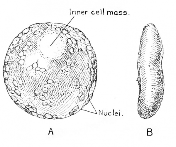
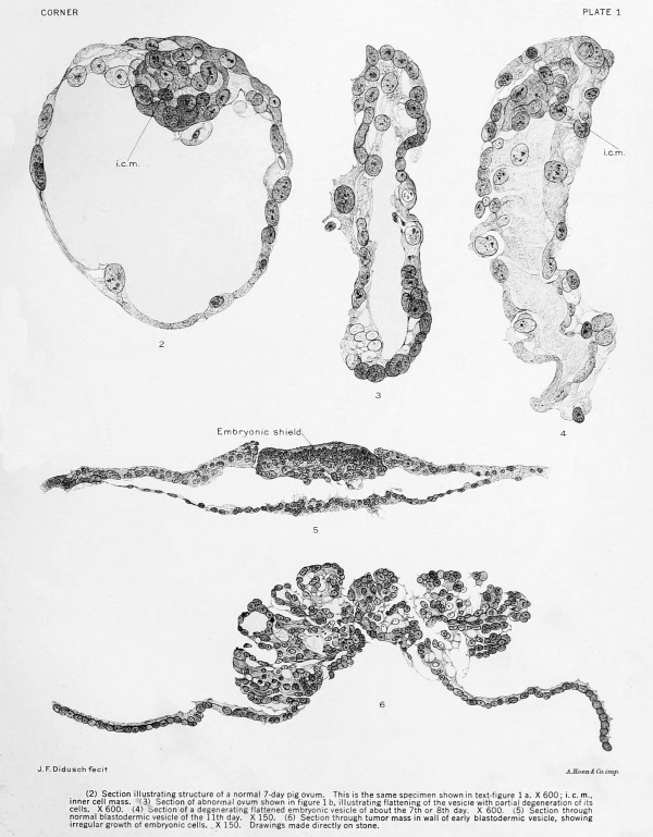
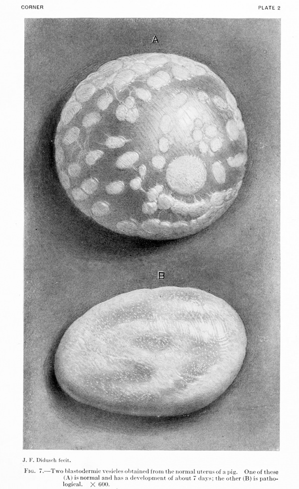
The specimens described herein were obtained during the collection of a series of early embryo pigs. Figure 1 shows two of these embryos that were found among others in a healthy, normal uterus. One (A) is a normal blastodermic vesicle which would appear, by comparison with Assheton's well-known stages, to be about 7 days old; figure 7a, plate 2, illustrating the same ovum, displays its delicate, filmy texture, with clearly outlined nuclei, and the developing embryonic area below the ectoblast. It is also shown in section in figure 2, plate 1. The other vesicle (b) is collapsed and wrinkled, its texture granular and almost opaque (the opacity is slightly exaggerated in fig. 7b), the nuclei hardly visible. Microscopic section confirms the external appearance of degeneration, showing the cells to be granular and their relations distorted (fig. 3). It is interesting to note that the preservation of the cells is better at one pole of the vesicle than at the other. In the ovaries of this sow there were 7 corpora lutca; in the uterus 6 vesicles, of which 2 were entirely normal, 2 normal in texture but collapsed and cup-shaped, and 2 abnormal, as illustrated. In addition there was 1 unsegmented ovum, thus accounting for all the ruptured follicles.
Figure 4 shows another case of the same sort with cupping of the vesicle and a partial break-down of the cells. This was obtained from another healthy uterus containing 1 normal vesicle and 4 of the collapsed type.
Fig. 1. A normal (A) and a pathological (B) ovum obtained from the same uterus. The more detailed structure of these specimens is shown in figure 7, plate 2. The pathological ovum is wrinkled and compressed. Here it is shown from a side view; in figure 7 its broader surface is shown.
A third sow contained a still more interesting series of 4 vesicles. These were 5 mm. in diameter, a size attained by normal pig embryos at about the twelfth day, according to the studies of Assheton. In 3 the abnormality consisted of total absence of an embryonic area; in order to make sure of this point the vesicles were fixed in the fully dilated condition by injection of Bouin's fluid. I am indebted to my colleague. Dr. C. H. Heuser, for friendly aid in the preparation of these specimens. When dilated the 3 vesicles were of even thickness at all points of their walls. Microscopic section of one of them showed it to be composed of the usual two layers of cells.
The fourth specimen from this uterus showed, on the contrary, a distinct mass, slightly larger than the embryonic area found in a normal vesicle of the same dimensions, but opaque, white, and projecting from the surface of the vesicle by a pedicle, so that altogether it was not unlike a little mushroom in appearance. Figure 6 shows a section of this curious specimen, in comparison with a normal embryo from a vesicle of the same size (fig. 5). The little tumor is composed of cells of diverse staining reaction, disposed in an irregular way, but including three or four minute vesicular cavities, one of which appears in the section. The whole arrangement rather suggests a malignant pai)illoma; whether the tumor is at the site of the embryonic area itself or at some other part of the vesicle (the embryonic area being absent, as in the other blastodermic vesicles in the same uterus) can not be decided ; but in any event the specimen may be regarded as a true monster, the carhest ever seen in a mammal. This and the other related blastodermic vesicles, like those described in the i:)revious paragraphs, were found lying unattached in the cavity of a healthy uterus which was quite normal and which proved on microscopic examination to be lined by a mucosa in every way similar to that of uteri containing normal embryos. The Fallopian tubes presented no gross abnormalities; they were not examined microscopically.
In view of the positive evidence afforded by the few specimens gathered by Huber and the writer, we are forced to the conclusion that the organism is liable to pathological changes before its attachment to the uterus. Whether these are due to faults inherent in the germ-cells, or to traumata which assail the ovum during its passage from the ovary to the uterine mucosa (such, for example, as chemically abnormal secretions of a seemingly normal uterus) our specimens of course do not explain; they merely increase the difficulty of the problem by demonstrating the inadequacy of the theory of faulty implantation to account for all developmental aberrations. Most hkely it will be proved in the end that the germ-cells and their product are liable to the onset of abnormalities at all stages of their historj^; certainly hints of such a possibihty are given by two lines of investigation, as yet but little applied to mammalian forms. The first of these comprises those experiments already mentioned, which show that the eggs of fish and frogs are susceptible to damage by all sorts of chemical and physical factors in their environment, at stages of development so early as to be comparable with the segmenting mammalian ovum during its passage through the tube and while in the uterus before implantation; the other line of approach is through the studies of Morgan and others, who have discovered the occurrence in plants, insects, and even mammals, of inheritable factors which, when brought into play by some unfavorable mating, cause such deviation from the anatomical or physiological norm as to kill the individual at one or another stage of its development. For instance, according to Cuenot, Castle, and Little, it is impossible to breed mice which are homozygous for yellow coatcolor; it appears that the inheritance of this character through both parents is invariably fatal to the embryo; and, indeed, the recent report of Kirkham hints strongly that in this case the lethal factor may attack the embryo shortly before the time of implantation, while it is still in the morula or vesicle stage. It seems not impossible, therefore, that in future the application to mammals of both the teratological and the genetic modes of experimentation may aid in solving the complex but pressing questions arising from abnormalities of development of the human organism.
Authors Cited
AssBETON, R., 189S. The development of the pig during the first ten days. Quart. Jour. Mierosc. Sci., vol. 41, p. 329.
Castle, W. E., and C. C. Little, 1910. On a modified Mendelian ratio among yellow mice. Science, n. 8., vol. 32, p. 868-870.
CrtNOT, L., 1908. Sur quclques anomalies apparentes des proportions Mend61ienne3. Arch. Zool. Exp., vol. 9; Notes et Revue II, p. vii.
HuDER, G. C, 1915. The development of the albino rat. II. Abnormal ova; end of the first to the end of the ninth day. Memoirs AVistar Inst. Anat. and Biol., No. 5.
Kellicott, AV. E., 1916. The effects of low temperature on the development of Fundulus. Amer. Jour. Anat., vol. 20, p. 449-482.
kinkham, W. B., 1917. Anat. Ree., Embryology of the yellow mouse, vol. 11, p. 480-481. (Proc. Amer. Soc. Zool.)
Mall, F. P., 1905. A study of the cause.? underlying the origin of human monsters. Jour. Morph., vol. 19. p. 1-361. , 1910.
- Pathology of the human ovum. In: Keibel and Mall. Manual of Human Embryology, vol. 1, p. 202-242. , 1917.
- On the frequency of localized anomalies in human embryos and infants at birth. Amer. Jour. Anat., vol. 22, p. 49-72.
Little, C. C, 1917. The relation of yellow coat color and black-eyed white spotting of mice in inheritance. Genetics, vol. 2, p. 433-444.
MoBQAN, T. H., 1917. The mechanism of Mendelian inheritance. New York. ,1919. The physical basis of heredity. Philadelphia.
Plates
Plate 1
Fig. 2. Section illustrating structure of a normal 7-day pig ovum. This is the same specimen shown in text-figure 1a. X600; i.c.m., inner ceil mass.
Fig. 3. Section of abnormal ovum shown in figure 1 b, illustrating flattening of the vesicle with partial degeneration of its cells. X 600.
Fig. 4. ejection of a degenerating flattened embryonic vesicle of about the 7th or 8th day. X 600.
Fig. 5. Section through normal blastodermic vesicle of the 1 1th day. X 150.
Fig. 6. Section through tumor mass in wall of early blastodermic vesicle, showmg irregular growth of embryonic cells. X 150. Drawings made directly on stone.
Plate 2
Fig. 7. Two blastodermic vesicles obtained from the normal uterus of a pig. One of these (A) is normal and has a development of about 7 days; the other (B) is pathological. X 600.
{{Historic Disclaimer}
Glossary Links
- Glossary: A | B | C | D | E | F | G | H | I | J | K | L | M | N | O | P | Q | R | S | T | U | V | W | X | Y | Z | Numbers | Symbols | Term Link
Cite this page: Hill, M.A. (2024, May 1) Embryology Book - Contributions to Embryology Carnegie Institution No.60. Retrieved from https://embryology.med.unsw.edu.au/embryology/index.php/Book_-_Contributions_to_Embryology_Carnegie_Institution_No.60
- © Dr Mark Hill 2024, UNSW Embryology ISBN: 978 0 7334 2609 4 - UNSW CRICOS Provider Code No. 00098G


