Book - Contributions to Embryology Carnegie Institution No.54
| Embryology - 27 Apr 2024 |
|---|
| Google Translate - select your language from the list shown below (this will open a new external page) |
|
العربية | català | 中文 | 中國傳統的 | français | Deutsche | עִברִית | हिंदी | bahasa Indonesia | italiano | 日本語 | 한국어 | မြန်မာ | Pilipino | Polskie | português | ਪੰਜਾਬੀ ਦੇ | Română | русский | Español | Swahili | Svensk | ไทย | Türkçe | اردو | ייִדיש | Tiếng Việt These external translations are automated and may not be accurate. (More? About Translations) |
Corner GW. A case of true lateral hermaphroditism in a pig with functional ovary. (1920) Contrib. Embryol., Carnegie Inst. Wash. Publ. , : 137-142.
| Historic Disclaimer - information about historic embryology pages |
|---|
 |
A Case of True Lateral Hermaphroditism in a Pig with Functional Ovary

By George W. Corner,
Of the Anatomical Laboratory, Johns Hopkins Medical School.
With one plate.
| Historic Disclaimer - information about historic embryology pages |
|---|
|
That variety of true hermaphroditism which is characterized by the presence
of an ovary on one side and a testis on the opposite side is one of the rarest forms
of structural abnormality of the genitalia. No undoubted cases have yet been
reported in man, and but two instances have been observed in swine. Some idea of
the rarity of the condition may be gained from the results of a systematic examination of 500,000 swine made in the Berlin municipal abattoir, under the direction
of Ludwig Pick (1914). In this series of animals five cases of hermaphroditism
were observed, but none of them was of the lateral variety, a mixed gland (ovotestis) on one or both sides being present in each.
Reuter (1885) described the case of a two months' old pig, discovered among
a litter which also contained two pseudohermaphrodites. This animal possessed a
right testis and a left ovary. The ovary was very small but contained ova in
primordial follicles; the testis contained numerous interstitial cells, but apparently
there were no germ-cells in the tubules. On the side on which the testis was located
the uterus ended in a rudimentary Fallopian tube.
Kingsbury (1909) recorded the examination of a young adult pig with male
external genitalia. There was a normal-looking uterus with a rudimentary left
ovary and a large right testis with a typical epididymis. On the right side the
Fallopian tube ended in a diminutive blind sac. The ovary contained a few ova
in follicles; the testis had the typical structure of a crytorchid testis, with numerous
interstitial cells but without germ-cells in the tubules.
Description of Specimen
The author's specimen, consisting of the uterus, tubes, and ovaries of an adult pig, was found among a number of uteri which had been brought in from a neighboring slaughter-house for study; therefore no information is at hand concerning the history or appearance of the animal from which it came.
The uterus was normally formed (fig. 1), presenting two cornua as usual. Its
size corresponded to that of the uterus of a young, sexually mature sow. On the
right side the uterine cornu ended in a normal Fallopian tube in connection with
a normal ovary; the latter contained four recent corpora lutea, one of them cystically dilated. On washing out the contents of the tube with saline solution, one
ovum was found, normal in all respects except that the cytoplasm was slightly
shrunken; one polar body had been extruded. The left uterine horn, normal in
size and form, ended in a very slender tube about 1 mm. in external diameter near the uterus, which gradually thinned down to an almost linear dimension, losing its
lumen, and finally ending in the connective tissue over the epididymis (fig. 1).
On the left side, in place of an ovary there was a mass 30 by 25 by 20 mm. in
diameter, of dull flesh-color, exactly resembling a testis in form, texture, and color.
It was covered by a thick capsule in which large and somewhat tortuous vessels
coursed; when this tunic was incised the contents bulged over the cut edges. The
exposed surface was dry and granular in appearance.
On this side of the uterus there was a well-defined Wolffian duct, such as is
occasionally present in sows, beginning in the vagina and running parallel to the
uterine horn between the layers of the broad ligament. However, instead of ending
in a cul-de-sac or in a series of minute cysts in the region of the ovarian pedicle, as
this duct usually does when present in the sow, it became greatly convoluted as it
approached the tip of the cornu and finally so closely coiled as to form the body
indicated in figure 1. This structure presented the appearance of an epididymis
by reason of its texture, its close apposition to the testis-like body, and also because
of a slight constriction at the middle portion, suggesting a division into globus major and minor.
Microscopic examination fully confirmed the foregoing interpretation and
proved that the specimen was indeed one of true lateral hermaphroditism. Sections of the testis (figs. 2 and 3) showed a typical tunica albuginea, within which
the gland substance consisted of tubules separated by relatively wide groups of
interstitial cells of normal appearance, in whose nuclei mitotic figures were occasionally found. The tubules were lined by a layer of high cells, nowhere more than
one cell deep, except that here and there a nucleus lay farther from the basal margin
than the others. The nuclei were of medium size and contained relatively less
chromatin than those of the interstitial cells. No mitoses could be found. The
cytoplasm toward the free border was frayed out into long irregular strands which
were so interlaced that the lumina of the tubules seemed in most places to be filled
by this vague network of protoplasmic material. Within the cell-bodies of this
epithelial lining there were numerous large vacuoles. Germ-cells were totally
lacking; no tubule contained any cells other than those already described, which
were presumably partially degenerated Sertoli cells. The connective tissue of the
testis was normal in appearance, showing no sign of the hyaline degeneration which
has sometimes been seen in hermaphrodite glands. The epididymis (fig. 4) was
similar in all respects to that of a normal male animal except that it contained no
spermatozoa.
In order to gain a general view of the ovary it was cut into six blocks and sections were taken from each of these portions; these presented everywhere the histological structure of a normally functioning organ (fig. 5) . The corpora lutea were
recently formed, with the membrana propria broken down and the elements of the
theca interna just beginning to invade the granulosa, indicating that ovulation had
taken place about three days before. (For grounds for this estimate, see Corner,
1919.) This finding is, of course, in accord with the presence of an ovum in the tube. No special significance is attached to the fact that one of the corpora lutea
was slightly cystic, since this is a common occurrence in normal swine. The cortex
of the ovary contained primordial ova and there were numerous follicles of normal
type. Due consideration was given to the possibility that small masses of testicular
tissue might be present in the ovary; that is, that the organ might be an ovotestis
(as in one of Pick's cases, winch macroscopically closely resembles ours), but no
foreign tissue was found.
The uterine mucosa was normal and similar in both horns. From the results
of studies on the cyclic changes in the uterine mucosa, now in preparation, the
author feels justified in stating that the uterus of this animal presented the microscopic features characteristic of the period of oestrus.
Discussion
Owing to the fortunate circumstance that this pig passed into the butcher's hands as a sexually mature animal just after an ovulation had occurred, we have had a unique opportunity to study the physiological state of the ovary; indeed, this is apparently the first sure case of glandular hermaphroditism in which there is direct evidence of the discharge into the deferent duct of germ-cells from either gonad.
In the presence of a normal ovary containing very early corpora lutea, an ovum in passage through the Fallopian tube, and a full-sized uterus histologically normal, it seems more simple to consider this animal as functionally a female (at least as far as the internal genitalia are concerned) in which a local malformation had substituted a functionless testis and an epididymis for one ovary and the corresponding oviduct. In this respect the conditions are much like those of the previously described examples of true hermaphroditism in swine (now numbering about fifteen), in each of which the internal genitalia have been feminine as to gross morphology, with a more or less well-developed uterus and a testis or ovotestis in the anatomical position of the ovary. The opposite type of hermaphroditism — the presence of an ovary or ovotestis at the usual site of the testis, with the genital duct system resembling the male type — should it occur, is far less likely to be observed by the anatomist because of the general custom of castration of boars intended for the butcher. In three of the four cases in man, however, which were summarized in the comprehensive review of L. Pick (1914), the hermaphrodite gland was found in the inguinal canal. The male gland of our specimen was not dissimilar to those of other cases. The close resemblance to the testes of ridglings, the absence of germ-cells, the relatively numerous interstitial cells, the remarkably complete epididymis, the persistent Wolffian duct, have all been commented upon in previous reports. So fully elaborated and indubitable a male apparatus as this must at once dispose of contentions such as that of Kermauner (1912), that the germ-lacking organs of supposed hermaphrodites are merely examples of nondifferentiation from a neutral state of the gonadal primordium. In view of the current debate as to the early history of the germ-cells in mammals, great interest attaches to the question as to whether these gamete-free testes of hermaphrodites at any time in their development contained spermatogonia ; and upon the answer to this depend the further questions as to why the ova seem always to survive at the expense of the spermatozoa, and in what manner the male germ-cells are inhibited; but these questions must await experimental attack or the chance discovery of embryonic stages of glandular hermaphroditism.
References Cited
Corner, G. Y., 1919. On the origin of the corpus luteum of the sow from both granulosa and theca interna. Am. Jour. Anat. 26, 117-183.
Kermatjnbr, F., 1912. Sexus anceps oder Hermaphroditismus. Frankf. Zeitschr. f. Pathol. 11.
Kincsuurt, B. F., 1909. Report of a case of hermaphroditism (H. verus lateralis) in Sus scrofa. Anatomical Record, 3, 27S-282.
Pick, L., 1914. Ueber den vvahren Hcrmaphroditismus des Menschen und der Saugetiere. Arch, f . mikr. Anat. 84, 2 Abt. 119-242.
Retjtek, J., 1885. Ein Beitrag zur Lehre von Hcrmaphroditismus. Verh. d. phys. med. Gesellsch. zu Wurzburg, N. F. xix.
Plate 1

(1) General view of specimen; X 0.75.
(2) Section of testis showing relative proportions of tubular and interstitial cells; X 28; hematoxylin and eosin.
(3) Section of testis showing details of structure; X 1300; iron hematoxylin.
(4) Section of epididymis showing normal character of tissue; X 80; hematoxylin and eosin.
(5) Section of ovary showing several small Graafian follicles and part of an early corpus luteum ; X 15; hematoxylin and eosin. These figures were drawn directly on stone from the gross specimen and from the sections.
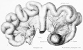
Figure 1
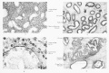
Figure 2 to 5

| Historic Disclaimer - information about historic embryology pages |
|---|
|
Glossary Links
- Glossary: A | B | C | D | E | F | G | H | I | J | K | L | M | N | O | P | Q | R | S | T | U | V | W | X | Y | Z | Numbers | Symbols | Term Link
Cite this page: Hill, M.A. (2024, April 27) Embryology Book - Contributions to Embryology Carnegie Institution No.54. Retrieved from https://embryology.med.unsw.edu.au/embryology/index.php/Book_-_Contributions_to_Embryology_Carnegie_Institution_No.54
- © Dr Mark Hill 2024, UNSW Embryology ISBN: 978 0 7334 2609 4 - UNSW CRICOS Provider Code No. 00098G