Book - An Atlas of Topographical Anatomy 5
V. Transverse section through the head
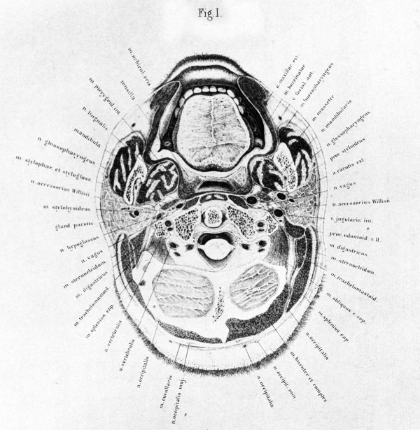
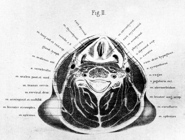
Male. Fig. 1. At the level of the teeth. Fig. 2. At the level of the upper edge of the thyroid cartilage and fifth cervical vertebra
| Embryology - 2 May 2024 |
|---|
| Google Translate - select your language from the list shown below (this will open a new external page) |
|
العربية | català | 中文 | 中國傳統的 | français | Deutsche | עִברִית | हिंदी | bahasa Indonesia | italiano | 日本語 | 한국어 | မြန်မာ | Pilipino | Polskie | português | ਪੰਜਾਬੀ ਦੇ | Română | русский | Español | Swahili | Svensk | ไทย | Türkçe | اردو | ייִדיש | Tiếng Việt These external translations are automated and may not be accurate. (More? About Translations) |
Braune W. An atlas of topographical anatomy after plane sections of frozen bodies. (1877) Trans. by Edward Bellamy. Philadelphia: Lindsay and Blakiston.
- Plates: 1. Male - Sagittal body | 2. Female - Sagittal body | 3. Obliquely transverse head | 4. Transverse internal ear | 5. Transverse head | 6. Transverse neck | 7. Transverse neck and shoulders | 8. Transverse level first dorsal vertebra | 9. Transverse thorax level of third dorsal vertebra | 10. Transverse level aortic arch and fourth dorsal vertebra | 11. Transverse level of the bulbus aortae and sixth dorsal vertebra | 12. Transverse level of mitral valve and eighth dorsal vertebra | 13. Transverse level of heart apex and ninth dorsal vertebra | 14. Transverse liver stomach spleen at level of eleventh dorsal vertebra | 15. Transverse pancreas and kidneys at level of L1 vertebra | 16. Transverse through transverse colon at level of intervertebral space between L3 L4 vertebra | 17. Transverse pelvis at level of head of thigh bone | 18. Transverse male pelvis | 19. knee and right foot | 20. Transverse thigh | 21. Transverse left thigh | 22. Transverse lower left thigh and knee | 23. Transverse upper and middle left leg | 24. Transverse lower left leg | 25. Male - Frontal thorax | 26. Elbow-joint hand and third finger | 27. Transverse left arm | 28. Transverse left fore-arm | 29. Sagittal female pregnancy | 30. Sagittal female pregnancy | 31. Sagittal female at term
| Historic Disclaimer - information about historic embryology pages |
|---|
| Pages where the terms "Historic" (textbooks, papers, people, recommendations) appear on this site, and sections within pages where this disclaimer appears, indicate that the content and scientific understanding are specific to the time of publication. This means that while some scientific descriptions are still accurate, the terminology and interpretation of the developmental mechanisms reflect the understanding at the time of original publication and those of the preceding periods, these terms, interpretations and recommendations may not reflect our current scientific understanding. (More? Embryology History | Historic Embryology Papers) |
THIS plate and Nos. 6, 7, and 8, are made from sections of one and the same body. The region of the neck was cut in five series of planes of which the upper surface of each is represented and analysed, it is viewed from above downwards. The right side of the drawing is the right side of the preparation. Owing to this cutting into planes, the explanation of the individual outlines becomes considerably more difficult than had the sections been made on different bodies. By making very thin sections the arrangement of the muscles of the nape of the neck were very difficult of definition. On the other hand, this proceeding affords the great advantage that the under surface of each section fits exactly on the upper surface of the one next following it, and also that the separate organs, such as the thyroid body and larynx, which show- pretty considerable individual differences with reference to size and position, can be analysed by transverse sections which mutually correspond. The body, which was of fine proportions and perfectly normal, was quite fresh, and was about twenty-five years of age. The muscular development was good. After the arteries had been injected, the trunk, from which the lower extremities were removed, was frozen in the usual manner, with the arms close to the side, and prepared as has been before described.
In consequence of the great muscular development the shoulders were very high, and therefore the neck appears to be comparatively short. It is not, then, to be wondered at that sections at the level of corresponding vertebrae in respect to the region of the shoulder, differ from those represented by Pirogoff (fasc. i, tab. ii), which were made from a person less thoroughly developed.
Fig. 1 corresponds nearly with Pirogoff (fasc. i, tab. Ix, fig. 1), and Henke (taf. Ixx, fig. 2). The section passes through the mouth and runs somewhat above the level of the teeth, falling upon the hard palate and the lateral masses of the first cervical vertebra, and slices off a thin lamina of the cerebellum on the posterior edge of the foramen magnum. It is seen on comparing it with Plate I that the section passes obliquely backwards and upwards,, from the head being bent somewhat backward in the recumbent position of the body. This relation must be borne in mind in observations on the living body. In the normal upright position of the body a plane section through the level of the teeth would pass through the second cervical vertebra, and would not touch the skull at all.
After cleansing the preparation it appeared that a small portion of the dorsum of the tongue also was sliced off with the crowns of the upper row of teeth. The apex of the tongue remained just behind the teeth. Posteriorly the section had passed 1'5 in. from the foramen caecum. The papillae which are seen on the posterior portion of the section correspond to the middle of the tongue. In the middle line from before backwards is the septum linguae, from which the fibres pass to both sides of the transverse muscles; in the posterior third the upper longitudinal fibres are seen. Behind the back of the tongue the uvula is retained in its entire length, as the section passed through the soft palate a quarter of an inch above its root, where the pillars of the fauces meet. The upper portion only of the tonsil is divided. In front of it, and behind the glands of the soft palate, lie some muscular fibres which pass transversely upwards, belonging to the upper border of the palato-glossus, which is embedded in the anterior pillar of the fauces. The azygos uvulae is also seen. Behind the tonsil and in relation with it is the palato-pharyngeus, which forms the posterior pillar of the fauces. The accurate division of the muscles cannot be defined, nevertheless it appears as if the transversely divided bundle of muscular fibre behind the tonsil (especially the left), belongs to the levator palati. There is no portion of the tensor palati seen since the section passed below the hamular process. A portion of the superior constrictor of the pharynx is very well shown in connection with the lower jaw and the buccinator.
Within this muscular zone is the cavity of the pharynx. This space is often thought to be larger because it is observed on the living body in an oblique direction, through the posterior palatine arch ; in a vertical section made on soft preparations the interval between the uvula and the posterior wall of the pharynx is generally represented as far too large. In the operation of staphyloraphy one is often disagreeably surprised at the narrowness of the locality, and must resort to some one of the ingenious needles which have been invented on account of this want of room.
Behind the muscular tissue of the pharynx and the lax cellular tissue which in the plate is represented as a white line, lie the longi colli and recti capitis antici majores, and further outwards on the transverse process of the atlas is the tendinous origin of the rectus capitis lateralis. The position of the internal carotid artery is of the utmost importance in operations on the tonsil and pharynx. It is seen that this large arterial trunk lies in immediate relation with the muscular tissue of the pharynx ; its pulsation can be easily felt from that cavity during life, and deep incisions in this region should not be made without the greatest caution.
The actual position of the artery to the tonsil, on the other hand, permits of greater freedom in extirpation of this gland, and numerous operations on it have shown that Hyrtl's apprehension (' Top. Anat.,' 1, 380) in this respect is much exaggerated. Nevertheless the proximity of the carotid must be especially borne in mind, even in the forcible dragging of the gland from its bed, but from the benign nature of most of the tumours of the tonsil there is no necessity for endeavouring to remove the gland completely, as the surgeon may be thoroughly satisfied if the chief mass of the growth be extirpated. As most of the instruments used for operations in this region only permit of a levelling and not of an extirpation of the tonsil, there is a sort of guarantee against wound of the carotid.* The position of the inferior dental and lingual nerves with regard to the lower jaw, is well shown. With regard to the latter nerve it is to be remarked that wounds of it in clumsy extraction of teeth from the slipping off of the instrument have often occurred. Its division in the mouth in neuralgia, as Eoser has recommended, is thoroughly practicable, without cutting through the cheek. After extraction of the last upper molar the nerve may be divided with a tenotome on the ramus of the jaw, without the necessity of further dissection. The articulation between the axis and atlas is such that the saw has entered under the anterior arch of the atlas, dividing its odontoid process, and has met the occipital bone over the posterior arch. The powerful transverse ligament of the atlas passing obliquely behind the odontoid process, is separated from the bone by a bursa. Further back is seen the broad ligamentous mass of the lateral axoid ligament, which terminates on the body of the axis, and which partly passes over into the posterior common ligament. I have been unable to find in this preparation the synovial membrane described by Luschka as existing between these ligaments. Unfortunately, the two ligaments are not clearly enough defined from each other in the plate ; the lateral portions of the ligamentum latum are represented too streaky. On the anterior aspect of the odontoid process lies the ligamentous mass filling up the space between the bodies of the axis and anterior arch of the atlas the deep anterior axo-atloid ligament. A portion of this ligament is met with below the anterior arch of the atlas ; the anterior articular cavity lies above the section. It will be seen from the breadth of the ligamentous mass that the position of the odontoid process acts as a safeguard against powerful strains, and that the lateral masses of the atlas must have a great expanse in order to afford sufficient attachments for such strong ligaments. The mass of cellular tissue which closes up the space between the posterior arch of the atlas and the occipital bone is very lax, and the posterior atlanto-occipital ligament, which is cut at a very acute angle, takes up a considerable space. Immediately beneath, the posterior arch of the atlas can be felt. It is here that the vertebral artery makes its way, in order to perforate the dura mater further internally, and it thus reaches the medulla oblongata. The artery is divided three times, on account of its curves. The first section is in the vertebral canal, where the artery passes vertically upwards, and the second where it bends backwards after the completion of the curve as an arc flattened transversely towards the middle line.
- By the use of a simple curved bistoury and vulsellum forceps the tonsil can be more readily and easily removed than by any other method, and the object of dragging the gland forcibly from its bed towards the mesial line, with a view of avoiding any chance of wounding the vessel, can be well recognised from examining the plate. TB.
There is nothing particular to observe with regard to the section of the skull and the small lamina of cerebellum. As the skull was divided very superficially the prominences appear larger than they really are, and they thus acquire such singular forms. The muscles, vessels, and nerves of this region are readily recognised in the plate, and require no particular remark. The occipital artery of the right side is seen through a considerable portion of its length. It arises from the posterior aspect of the external carotid, and passing at first vertically upwards, crosses the internal jugular vein to reach the posterior belly of the digastric. From thence it runs horizontally backwards in the lateral region of the neck, being covered by the trachelo-mastoid and splenius. Having arrived at the middle edge of the splenius it pierces the upper origin of the trapezius (cucullaris), and then runs superficially on the skull. Very little of this vessel is to be seen on the left side. Between the splenius and the occipital bone a muscular branch appears passing deeply from its trunk.
The glosso-pharyngeal, vagus, spinal accessory, and hypoglossal nerves are shown in the plate.
The parotid gland is of especial surgical interest ; it is enclosed in a dense fascial envelope which surrounds it on all sides, sending a multitude of septa into the substance of the gland, which account for the tabulated appearance which it presents on section. As the fascia lines the entire niche in which the parotid is imbedded, there is not only a demarcation between it and the internal jugular vein (which must especially be considered in the extirpation of tumours of the gland), but it is a protection to the vagus, spinal accessory, and hypoglossal nerves, which are in close proximity. The portion of the fascia which is most strongly developed is that which covers in the outer aspect of the gland. This, on account of its connection with the fascia of the masseter, is called fascia rnasseterico-parotidea.
In consequence of this arrangement the swelling of the gland from inflammation is limited externally, thus the tumour presses internally against the nerves and vessels.
The parotid being pierced by the terminal branches of the external carotid artery and the posterior facial vein, its extirpation without wounding these vessels is impracticable. But if the carotid, as shown on the right side of this preparation, lies so peripherally that it can be dug out of the mass of gland tissue, an operation would be less uncretain in its result.
On account of the numerous anastomoses of the arteries in the skull it is of little use to attempt to arrest haemorrhage from the external carotid, but in the event of complete extirpation of the gland it would be necessary to direct one's attention especially to the preservation of the internal jugular vein.
Figure 2 represents the upper surface of a lamina 1*5 inch thick, which corresponds with the under surface of Plate IV. The section which the plate represents has passed through the thyroid notch and has fallen close on the upper border of the fifth cervical vertebra.
As the section passed immediately below the chin and lower jaw, the neck would be divided in its so-called cylindrical portion. It is seen, however, that on account of the muscular development at this level the natural form of the neck is not an exact cylinder, consequently its section is not a circle, but is a pentagon.
The lateral portion of the trapezius (cucullaris) commences immediately below the section, consequently the plane of section is enlarged, corresponding with the anterior curvature of the cervical spine. The section of the vertebra lies much further removed from the side of the neck than one would expect. In the accompanying plate the body of the vertebra lies in the anterior half of the figure. The point met with in the section of the vertebra is where the arch springs from the body, hence the lumen of the spinal canal is seen. On the left side is seen the articular process of the sixth cervical vertebra, and from this point we can follow the course of the sixth cervical nerve behind and. to the outer side of the vertebral artery. The divided nerve seen lying in the bifurcation of the transverse process is the fifth cervical.
The larynx is so divided that the vocal cords with the ventricle of Morgagni between them is clearly shown. The mucous membrane which lies behind the divided arytenoid cartilages is here singularly rich in glands.
In the section also are seen the arytenoid glands, many of which are embedded on the inner side of the aryteno-epiglottidean fold. The thyro-arytenoideus and the arytenoideus are divided through their upper extremities. From the arrangement of the muscles, the resemblance to a sphincter can be clearly recognised. Behind this layer of muscles lies the large mass of glands of the pharynx the middle arytenoid gland of Luschka.
The common carotid artery is shown exactly in the position which would be most suitable for its ligature ; its relations should be carefully noticed. It is at the spot where the deviation of the omo-hyoid and sterno-cleido-mastoid allows of a ready means of access.
However incompletely the relations of the fasciae in such a plate are rendered, one can see clearly that the vessel must be sought on the anterior border of the sterno-cleido-mastoid, and that after the division of the hinder portion of its sheath the space containing the great vessels and nerves is immediately entered.
In front of the artery is the descendens noni, and somewhat behind it is the internal jugular vein between the vein and artery is the vagus, and behind the artery is the sympathetic. Inside the sheath of the vessels a layer of cellular tissue isolates the artery from the vein and the nerve. The important part of the operation consists in opening the sheath of the artery, which lies immediately in front of the scalenus anticus muscle. If this be properly done, one not only avoids the danger of wounding the nerve, but the vein also will be kept at a distance. After opening the sheath the vein would expand enormously and cover up the whole field of operation.*
- In ligature of the carotid pressure should be made on the internal jugular vein by the fingers of an assistant both above and below, in order to prevent this expansion of the vessel from interfering with the operator's movements. TB.
| Historic Disclaimer - information about historic embryology pages |
|---|
| Pages where the terms "Historic" (textbooks, papers, people, recommendations) appear on this site, and sections within pages where this disclaimer appears, indicate that the content and scientific understanding are specific to the time of publication. This means that while some scientific descriptions are still accurate, the terminology and interpretation of the developmental mechanisms reflect the understanding at the time of original publication and those of the preceding periods, these terms, interpretations and recommendations may not reflect our current scientific understanding. (More? Embryology History | Historic Embryology Papers) |
- Braune Plates (1877): 1. Male - Sagittal body | 2. Female - Sagittal body | 3. Obliquely transverse head | 4. Transverse internal ear | 5. Transverse head | 6. Transverse neck | 7. Transverse neck and shoulders | 8. Transverse level first dorsal vertebra | 9. Transverse thorax level of third dorsal vertebra | 10. Transverse level aortic arch and fourth dorsal vertebra | 11. Transverse level of the bulbus aortae and sixth dorsal vertebra | 12. Transverse level of mitral valve and eighth dorsal vertebra | 13. Transverse level of heart apex and ninth dorsal vertebra | 14. Transverse liver stomach spleen at level of eleventh dorsal vertebra | 15. Transverse pancreas and kidneys at level of L1 vertebra | 16. Transverse through transverse colon at level of intervertebral space between L3 L4 vertebra | 17. Transverse pelvis at level of head of thigh bone | 18. Transverse male pelvis | 19. knee and right foot | 20. Transverse thigh | 21. Transverse left thigh | 22. Transverse lower left thigh and knee | 23. Transverse upper and middle left leg | 24. Transverse lower left leg | 25. Male - Frontal thorax | 26. Elbow-joint hand and third finger | 27. Transverse left arm | 28. Transverse left fore-arm | 29. Sagittal female pregnancy | 30. Sagittal female pregnancy | 31. Sagittal female at term
Reference
Braune W. An atlas of topographical anatomy after plane sections of frozen bodies. (1877) Trans. by Edward Bellamy. Philadelphia: Lindsay and Blakiston.
Glossary Links
- Glossary: A | B | C | D | E | F | G | H | I | J | K | L | M | N | O | P | Q | R | S | T | U | V | W | X | Y | Z | Numbers | Symbols | Term Link
Cite this page: Hill, M.A. (2024, May 2) Embryology Book - An Atlas of Topographical Anatomy 5. Retrieved from https://embryology.med.unsw.edu.au/embryology/index.php/Book_-_An_Atlas_of_Topographical_Anatomy_5
- © Dr Mark Hill 2024, UNSW Embryology ISBN: 978 0 7334 2609 4 - UNSW CRICOS Provider Code No. 00098G


