Book - An Atlas of Topographical Anatomy 31
| Embryology - 28 Apr 2024 |
|---|
| Google Translate - select your language from the list shown below (this will open a new external page) |
|
العربية | català | 中文 | 中國傳統的 | français | Deutsche | עִברִית | हिंदी | bahasa Indonesia | italiano | 日本語 | 한국어 | မြန်မာ | Pilipino | Polskie | português | ਪੰਜਾਬੀ ਦੇ | Română | русский | Español | Swahili | Svensk | ไทย | Türkçe | اردو | ייִדיש | Tiếng Việt These external translations are automated and may not be accurate. (More? About Translations) |
Braune W. An atlas of topographical anatomy after plane sections of frozen bodies. (1877) Trans. by Edward Bellamy. Philadelphia: Lindsay and Blakiston.
- Plates: 1. Male - Sagittal body | 2. Female - Sagittal body | 3. Obliquely transverse head | 4. Transverse internal ear | 5. Transverse head | 6. Transverse neck | 7. Transverse neck and shoulders | 8. Transverse level first dorsal vertebra | 9. Transverse thorax level of third dorsal vertebra | 10. Transverse level aortic arch and fourth dorsal vertebra | 11. Transverse level of the bulbus aortae and sixth dorsal vertebra | 12. Transverse level of mitral valve and eighth dorsal vertebra | 13. Transverse level of heart apex and ninth dorsal vertebra | 14. Transverse liver stomach spleen at level of eleventh dorsal vertebra | 15. Transverse pancreas and kidneys at level of L1 vertebra | 16. Transverse through transverse colon at level of intervertebral space between L3 L4 vertebra | 17. Transverse pelvis at level of head of thigh bone | 18. Transverse male pelvis | 19. knee and right foot | 20. Transverse thigh | 21. Transverse left thigh | 22. Transverse lower left thigh and knee | 23. Transverse upper and middle left leg | 24. Transverse lower left leg | 25. Male - Frontal thorax | 26. Elbow-joint hand and third finger | 27. Transverse left arm | 28. Transverse left fore-arm | 29. Sagittal female pregnancy | 30. Sagittal female pregnancy | 31. Sagittal female at term
| Historic Disclaimer - information about historic embryology pages |
|---|
 |

THE body from which this plate was made, was that of a person thirty- five years of age, who died from drink at the commencement of labour. An examination of the genitals showed that the liquor amnii had not escaped. After having been prepared in the usual way, a section was made in the mesial plane from below upwards. The symphysis was not however exactly divided at its centre, but the deviation was so slight that it need not be regarded.
After the drawings were made of the right half of the body, and completely finished, the maternal structures were removed, in order to obtain the other half of the child uninjured and in its original position.
The child was a well-formed male of about six pounds weight including the cord which passed downwards under the left leg, whence it was bent upwards and lay over the left ankle joint, being reflected sharply on to the placenta which was attached to the upper portion of the uterus. The cord must have been cut through on removing the left half of the child, as I afterwards found its placental insertion in the left half of the body. I had divided it close to its placental extremity, and it was so firmly pressed against the child, that it could be with difficulty removed without inducing a change in the position of the left lower extremity.
The child's head as is seen in the plate is apparently in the second position, and was on the point of being born at the death of the mother. The natural rotation of the head in the pelvis has commenced, being
As this chapter refers almost entirely to the section of the child, and the corresponding plates are not reproduced in this small edition, I have thought it advisable to omit such portions of it as are not illustrated directly to the accompanying plate, and to advise the reader interested in the matter to consult Prof. Braune's ' Die Lage des Uterus und Fretus am Ende der Schwangerschaft,' which has been already translated into English. TE.
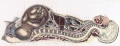
Uterus and Fetus Position at Birth
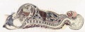
Section through Uterus and Fetus at Birth
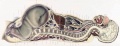
Uterus without Fetus at Birth
| Historic Disclaimer - information about historic embryology pages |
|---|
|
- Braune Plates (1877): 1. Male - Sagittal body | 2. Female - Sagittal body | 3. Obliquely transverse head | 4. Transverse internal ear | 5. Transverse head | 6. Transverse neck | 7. Transverse neck and shoulders | 8. Transverse level first dorsal vertebra | 9. Transverse thorax level of third dorsal vertebra | 10. Transverse level aortic arch and fourth dorsal vertebra | 11. Transverse level of the bulbus aortae and sixth dorsal vertebra | 12. Transverse level of mitral valve and eighth dorsal vertebra | 13. Transverse level of heart apex and ninth dorsal vertebra | 14. Transverse liver stomach spleen at level of eleventh dorsal vertebra | 15. Transverse pancreas and kidneys at level of L1 vertebra | 16. Transverse through transverse colon at level of intervertebral space between L3 L4 vertebra | 17. Transverse pelvis at level of head of thigh bone | 18. Transverse male pelvis | 19. knee and right foot | 20. Transverse thigh | 21. Transverse left thigh | 22. Transverse lower left thigh and knee | 23. Transverse upper and middle left leg | 24. Transverse lower left leg | 25. Male - Frontal thorax | 26. Elbow-joint hand and third finger | 27. Transverse left arm | 28. Transverse left fore-arm | 29. Sagittal female pregnancy | 30. Sagittal female pregnancy | 31. Sagittal female at term
Reference
Braune W. An atlas of topographical anatomy after plane sections of frozen bodies. (1877) Trans. by Edward Bellamy. Philadelphia: Lindsay and Blakiston.
Glossary Links
- Glossary: A | B | C | D | E | F | G | H | I | J | K | L | M | N | O | P | Q | R | S | T | U | V | W | X | Y | Z | Numbers | Symbols | Term Link
Cite this page: Hill, M.A. (2024, April 28) Embryology Book - An Atlas of Topographical Anatomy 31. Retrieved from https://embryology.med.unsw.edu.au/embryology/index.php/Book_-_An_Atlas_of_Topographical_Anatomy_31
- © Dr Mark Hill 2024, UNSW Embryology ISBN: 978 0 7334 2609 4 - UNSW CRICOS Provider Code No. 00098G