Book - An Atlas of Topographical Anatomy (1877)
| Embryology - 30 Apr 2024 |
|---|
| Google Translate - select your language from the list shown below (this will open a new external page) |
|
العربية | català | 中文 | 中國傳統的 | français | Deutsche | עִברִית | हिंदी | bahasa Indonesia | italiano | 日本語 | 한국어 | မြန်မာ | Pilipino | Polskie | português | ਪੰਜਾਬੀ ਦੇ | Română | русский | Español | Swahili | Svensk | ไทย | Türkçe | اردو | ייִדיש | Tiếng Việt These external translations are automated and may not be accurate. (More? About Translations) |
Braune W. An atlas of topographical anatomy after plane sections of frozen bodies. (1877) Trans. by Edward Bellamy. Philadelphia: Lindsay and Blakiston.
| Online Editor |
|---|

|
| Historic Disclaimer - information about historic embryology pages |
|---|
|

An Atlas of Topographical Anatomy After Plane Sections Of Frozen Bodies
By
Wilhelm Braune
Professor Of Anatomy In The University Of Leipzig
With Forty-Six Woodcuts In The Text
Translated By
Edward Bellamy, F.B.C.S.
Senior Assistant Surgeon To The Charing Cross Hospital; Lecturer On Anatomy And Teacher Of
Operative Surgery In Its School; Professor Of Anatomy As Applied To The Fine
Arts In The Science And Art Department, South Kensington
Philadelphia
Lindsay And Blakiston 1877
To
Christopher Heath, F.R.C.S.
Surgeon To University College Hospital And Holme Professor Of Clinical Surgery
In University College The English Edition Of This Work Is Inscribed In Recognition Of The Estimation In Which His Professional Skill Is, From Personal Experience, Held The Translator
Authors Preface
THE accompanying plates, representing plane sections of the human body, are reproduced on a smaller scale from my large atlas, in which the figures are the size of nature ; and are intended to assist in extending and increasing the knowledge of the human form, and of the position of the different organs to each other. The necessity for such plates for the clinic has repeatedly been expressed, and especially for military surgery. It has been emphatically stated on all sides that the sections of the thorax and abdomen are indispensable for diagnosis, and that the examination of my plates has been of substantial assistance in judging correctly the direction of a gunshot wound. Consequently when the question arose as to preparing another edition of the large coloured atlas, it seemed advisable at the same time to arrange a smaller one, which could be made accessible to a wider circle of readers. The plates of the large atlas are faithfully reproduced by photography. Some few plates have been omitted as of subordinate interest. The text remains unaltered, with the exception of a few additions.
As each plate has its special text appended, a detailed use of the atlas is rendered possible ; and, as circumstances require, recapitulations are introduced for the elucidation of individual plates. In like manner the precise data relating to the structure of the individual subjects are retained and repeated in connection with the plates, in order that the observer, by examining them, may avoid mistakes on the living body.
It will be perceived that the work is not intended to be a text-book of topographical anatomy, but an atlas, which may take its place amongst manuals on the subject, as an illustrated means of assistance.
WILH. BRAUNE.
November, 1874.
THE great success of Professor Braune's Atlas abroad has induced him to publish a smaller edition of his large work, with photographs of the original plates reduced to half-scale. It has been considered advisable to take advantage of this to reproduce the volume in English.
The immense expense of producing such plates and the persistent dearth of material have, in all probability, been the cause why no original English work on topographical anatomy has as yet been placed within the reach of the generality of students.
It is, I think, generally admitted that there is a want in this country of a good text-book on applied anatomy, and not a mere handbook, but such a work as might take its place with those of Richet, Hyrtl, or Luschka. By means of the sections found in this Atlas the exact position and relations of the structures which must be divided or avoided in the course of an operation are indicated ; and the track of a bullet or punctured wound suggested. At the same time they afford an absolutely correct representation of the intimate relations of the viscera of the thorax and abdomen.
I cannot help thinking that the work may be of great value to artists, as demonstrating the exact position of the bones to the muscles and indicating the contours of the body.
I have endeavoured to avoid a slavish translation of the text, and to reproduce the author's meaning in readable English, without interfering more than was absolutely necessary with the original construction of the sentences. I have taken the liberty of omitting some irrelevant matter, such as the repetition of methods of preparation, &c., which would be unnecessary and burdensome to the English student. In the text to Plates XXIX (A, B), XXX and XXXI, finding that the description of the section of the foetus referred to the author's large work more particularly and was not in any way illustrated by the present series of plates, I omitted it. I trust that Professor Braune will not consider that I have in any way mutilated his text or impaired its utility.
I cannot of course hold myself answerable for the opinions of the author in his surgical and medical ' comments, but have simply rendered ' them as I trust he intends them to be understood. I have reduced the measurements to their equivalent English notation (with the exception of the long table on page 155), on the advice of friends, although I consider that it would have been, in some respects, preferable to have retained the metric scale.
I must express my warmest thanks to my friends, Mr. Edmund B. Owen, F.R.C.S., Lecturer on Anatomy in St. Mary's Hospital, and Dr. J. Mitchell Bruce, M.A., Assistant-Physician to Charing Cross Hospital, for their kindness in revising the proof-sheets, and for many valuable hints.
EDWARD BELLAMY.
MARGARET STREET, CAVENDISH SQUARE ; October, 1876.
Contents
- Sagittal section of the body of a male, set.
- Sagittal section of the body of a female, set.
- Obliquely transverse section of the head, passing through the eyeballs ; female, set.
- Transverse section through the internal ear ; male
- Transverse section through the head; male. Fig. 1. At the level of the teeth. Fig. 2. At the level of the upper edge of the thyroid cartilage and fifth cervical vertebra
- Transverse section of the same body through the neck at the level of the cricoid cartilage and sixth cervical vertebra.
- Transverse section of the same body through the neck and shoulders at the level of the seventh cervical vertebra.
- Transverse section of the same body through the apices of the lungs and shoulderjoints at the level of the first dorsal vertebra.
- Transverse section of the thorax of a male at the level of the third dorsal vertebra.
- Transverse section of the same body at the level of the arch of the aorta and fourth dorsal vertebra.
- Transverse section of the same body at the level of the bulbus aortse and sixth dorsal vertebra.
- Transverse section of the same body at the level of the mitral valve and eighth dorsal vertebra.
- Transverse section of the same body at the level of the apex of the heart and ninth dorsal vertebra.
- Transverse section of the same body through the liver, stomach, and spleen, at the level of the eleventh dorsal vertebra.
- Transverse section of the same body through the pancreas and kidneys at the level of the first lumbar vertebra.
- Transverse section of the same body through the transverse colon at the level of the umbilicus and intervertebral space between the third and fourth lumbar vertebra.
- Transverse section of the same body through the pelvis at the level of the upper portion of the head of the thigh bone.
- Transverse section through the pelvis of a male, set. 25, through the lower portion of the head of the thigh bone.
- Fig. 1. Vertical section of an injected knee-joint; female, middle age. Fig. 2. Vertical section through the right foot, close to its inner edge, from the same body.
- Fig. 1. Transverse section through the upper portion of the thigh, parallel with and close to Poupart's ligament (same body as Plate I). Fig. 2. Transverse section through the left thigh of the same body close to the trochanter minor.
- Fig. 1. Transverse section of the left thigh, just below the middle third, from the same body. Fig. 2. Transverse section of the left thigh through the middle, from the same body.
- Fig. 1. Transverse section of the lower third of the left thigh (male, middle age). Fig. 2. Transverse section through the left knee of the same body.
- Fig. 1. Transverse section through the upper third of the left leg of the same body. Fig. 2. Transverse section through the middle of the left leg of the same body.
- Fig. 1. Transverse section through the lower third of the left leg of the the same body. Fig. 2. Transverse section through the malleoli of the same.
- Frontal section through the thorax ; male.
- Fig. 1. Vertical section through the right elbow-joint ; female. Fig. 2. Vertical section through the hand and third finger of the same body.
- Figs. 1-4. Transverse section through the left arm, through the middle of the lower third of the humerus, through the trochlea, and head of the radius ; male, set.
- Figs. 1-4. Transverse section through the left fore-arm of the same, the upper middle and lower thirds, and wrist -joint.
- Sagittal sections through the body of a female in advanced pregnancy.
- Sagittal sections through the body of a female in advanced pregnancy.
- Sagittal sections through the lower half of a female at full term.
Plate Gallery
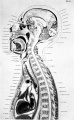
Plate 1A
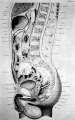
Plate 1B
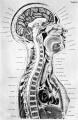
Plate 2A
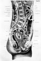
Plate 2B
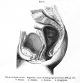
Plate 2 Fig.1
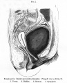
Plate 2 Fig.2
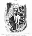
Plate 2 Fig.3
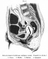
Plate 2 Fig.4
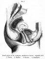
Plate 2 Fig.5
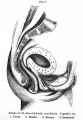
Plate 2 Fig.6

Plate 2 Fig.7
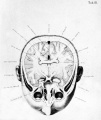
Plate 3
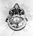
Plate 4
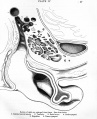
Plate 4 Fig.1
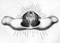
Plate 6
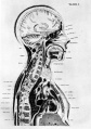
Plate 29A
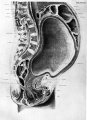
Plate 29B
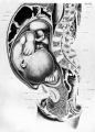
Plate 30

Plate 30 Fig.1
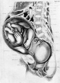
Plate 31
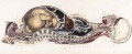
Plate 29 and 30. Uterus and Fetus Position at Term
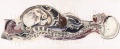
Plate 29 and 30. Section through Uterus and Fetus
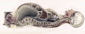
Plate 29 and 30. Uterus without Fetus
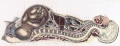
Plate 31. Uterus and Fetus Position at Birth
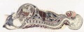
Plate 31. Section through Uterus and Fetus
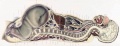
Plate 31. Uterus without Fetus


| Historic Disclaimer - information about historic embryology pages |
|---|
|
- Braune Plates (1877): 1. Male - Sagittal body | 2. Female - Sagittal body | 3. Obliquely transverse head | 4. Transverse internal ear | 5. Transverse head | 6. Transverse neck | 7. Transverse neck and shoulders | 8. Transverse level first dorsal vertebra | 9. Transverse thorax level of third dorsal vertebra | 10. Transverse level aortic arch and fourth dorsal vertebra | 11. Transverse level of the bulbus aortae and sixth dorsal vertebra | 12. Transverse level of mitral valve and eighth dorsal vertebra | 13. Transverse level of heart apex and ninth dorsal vertebra | 14. Transverse liver stomach spleen at level of eleventh dorsal vertebra | 15. Transverse pancreas and kidneys at level of L1 vertebra | 16. Transverse through transverse colon at level of intervertebral space between L3 L4 vertebra | 17. Transverse pelvis at level of head of thigh bone | 18. Transverse male pelvis | 19. knee and right foot | 20. Transverse thigh | 21. Transverse left thigh | 22. Transverse lower left thigh and knee | 23. Transverse upper and middle left leg | 24. Transverse lower left leg | 25. Male - Frontal thorax | 26. Elbow-joint hand and third finger | 27. Transverse left arm | 28. Transverse left fore-arm | 29. Sagittal female pregnancy | 30. Sagittal female pregnancy | 31. Sagittal female at term
Reference
Braune W. An atlas of topographical anatomy after plane sections of frozen bodies. (1877) Trans. by Edward Bellamy. Philadelphia: Lindsay and Blakiston.
Glossary Links
- Glossary: A | B | C | D | E | F | G | H | I | J | K | L | M | N | O | P | Q | R | S | T | U | V | W | X | Y | Z | Numbers | Symbols | Term Link
Cite this page: Hill, M.A. (2024, April 30) Embryology Book - An Atlas of Topographical Anatomy (1877). Retrieved from https://embryology.med.unsw.edu.au/embryology/index.php/Book_-_An_Atlas_of_Topographical_Anatomy_(1877)
- © Dr Mark Hill 2024, UNSW Embryology ISBN: 978 0 7334 2609 4 - UNSW CRICOS Provider Code No. 00098G