Advanced - Heart Tubes
| Begin Advanced | Heart Fields | Heart Tubes | Cardiac Looping | Cardiac Septation | Outflow Tract | Valve Development | Cardiac Conduction | Cardiac Abnormalities | Molecular Development |
| Cardiac Embryology | Begin Basic | Begin Intermediate | Begin Advanced |
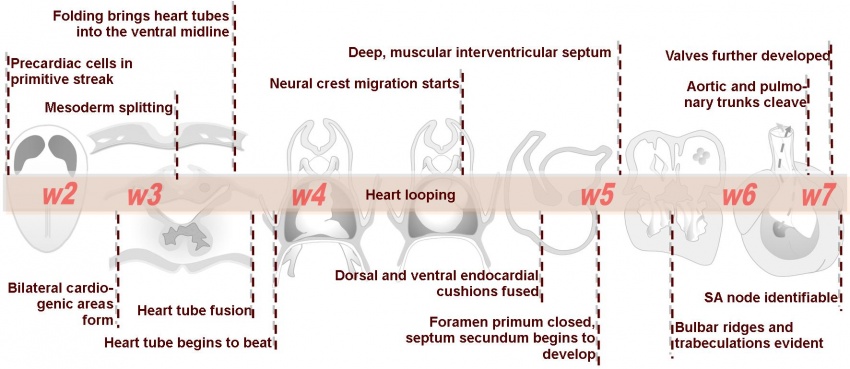
At approximately day 19 the lateral plate mesoderm divides into dorsal (somatic) and ventral (splanchnic) layers, forming the pericardial coelom between them. Angioblastic cords develop in the splanchnic mesoderm and canalise to form bilateral heart tubes. Following lateral folding of the embryo, fusion of the heart tubes occurs, beginning cranially and extending caudally. Folding of the heart of the embryo during the fourth week brings the heart tube dorsal to the pericardial cavity. The precardiomyocytes differentiate to form the myocardial sleeve of the heart tube, allowing the heart tube to begin beating. By the end of the fourth week (day 22) coordinated contractions of the heart tube, which push blood cranially, are present. This function initiates as nutritional and oxygen embryologic demands can no longer be met by passive diffusion from the placenta.
The primordial myocardium forms from splanchnic mesoderm surrounding the pericardial coelom. It is separated from the endothelial heart tube by cardiac jelly (gelatinous connective tissue). The endothelium of the heart tube forms the internal endocardium, while the epicardium develops from mesothelial cells arising from the sinus venosus, which spread cranially over the myocardium. Recent knowledge shows that additional myocardial cells are added to the outflow tract during heart looping. The following animation shows the folding and fusion of the heart tubes (Click image to play on current page or Play video on new page).
<html5media height="720" width="560">File:Heart_folding_001.mp4</html5media>
| Back to Heart Fields | Next: Cardiac Looping | |
| Go to this section in the intermediate level |
