2011 Lab 5 - Fetal
| 2011 Lab 5: Introduction | Trilaminar Embryo | Early Embryo | Late Embryo | Fetal | Postnatal | Abnormalities | Quiz | Online Assessment |
Fetal Development
The Embryonic period involved transient structures to establish body and placental tissues, folding and form. The long human Fetal period (4x the embryonic period) is a time of extensive growth in size and mass as well as ongoing differentiation of organ systems established in the embryonic period. (More? See Fetal Notes)
As many students are new to embryology the majority of the practical class time has been spent introducing general concepts of early human development and combining this with the early formation of GIT structures. While the fetal period is substantially longer, there will be much less content to be covered.
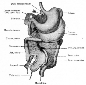
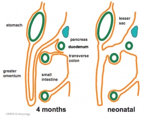
Fetal developmental features include: the growth and rotation of intestines initially herniated outside the ventral body wall; changes in mesenteries; development of the blood supply and tract wall.
Finally consider the initial functions of the tract with amionic fluid swallowing and the accumulation of both secretions and swallowed components within the large intestine as meconium.
Early Fetus
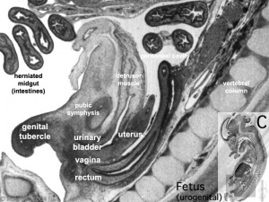
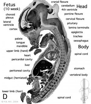
Begin by just looking at the fetal anatomy in sections taken through the above 10 week female fetus (which is approximately 40 mm in size) and see how this compares with what you already know of adult GIT anatomy.
Related Images
Fetus (week 10) Planes A (most lateral), B (lateral), C (medial) and D (midline) from lateral towards the midline.
- Human Fetus - most lateral | lateral | medial | midline
- Head - most lateral | lateral | medial | midline
- Cerebellum - most lateral | lateral | medial | midline
- Urogenital Unlabelled - most lateral | lateral | medial | midline
- Urogenital Labelled - most lateral | lateral | medial | midline
- Large Images - midline
- Image Source: UNSW Embryology, no reproduction without permission.
There are 4 sections taken in the parasagittal and sagittal plane (moving from the right at Plane A towards the midline at Plane D). Click on the small images (or the text below) to open the linked large image pages.
Midgut Herniation
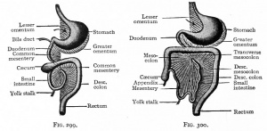
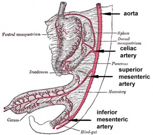
In the week 10 fetus (right) the midgut is still herniated (lying outside the ventral body wall) at the umbilicus. We saw in earlier pages that this heriation began back in the embryonic period (week 5) and the initial loop undergoes a series of rotations through embryonic and early fetal periods which position the midgut in its correct adult anatomical locations. These rotations are around the superior mesenteric artery (which supplies this region) and the associated mesentry. (Note this rotation is complicated and explained differently in different texts)
One week later (week 11) continued fetal body wall growth (and other factors) returns the midgut herniation returns to the abdominal cavity.
Small intestine growth in length is initially linear (first half pregnancy to 32 cm CRL), followed by rapid growth in the last 15 weeks doubling the overall length to term. Growth continues postnatally but after 1 year slows again to a linear increase to adulthood. (More? Intestine Development)
| <Flowplayer height="265" width="320" autoplay="true">Midgut_rotation_cartoon.flv</Flowplayer> | View from left hand side of embryo facing left. Sequence spans approx 6-11 weeks of development.
Quicktime movie | Quicktime | [[Development_Animation_-_Midgut_rotation|Flash] |
Stomach Mesentery
Blood Supply
|
Each region of the gastrointestinal tract has a specific arterial supply which historically defined the 3 gastrointestinal tract regions (foregut, midgut and hindgut).
|
Fetal Tract Development
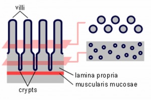
Week 11 - villi begin to appear in small intestine, goblet cells present
Week 16 - villi apparent in entire intestine
Adult small intestine will be divided into 3 regions with the same basic histological organization.
- duodenum (25-30 cm)
- jejunum (about first two-fifths of the rest)
- ileum
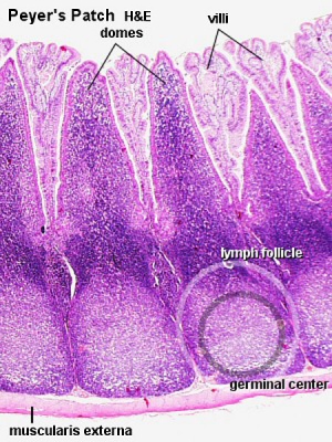
Week 20 - Peyer's patches appear in small intestine
The adult intestinal immune system includes:
- Peyer's patches
- isolated lymphoid follicles
- cryptopatches - small clusters of lymphoid cells with an immature lymphocyte phenotype and dendritic cells.
- mesenteric lymph nodes
Amniotic Fluid and Meconium
Amniotic Fluid Swallowing In early embryonic development both the buccopharyngeal and cloacal membranes degenerated, allowing the GIT to be filled with amniotic fluid. Towards the end of the fetal period the fetus is now swallowing approximately 500 ml of amniotic fluid / day.
This swallowed amniotic fluid moves through the GIT from esophagus, to stomach, to small intestine, stopping at the large bowel. In the large bowel the majority of fluid (water) is absorbed, along with electrolytes, glucose, urea and hormones. This process may contribute to fetal nutrition and prepare the GIT for its postnatal function. The process of swallowing amniotic fluid has been suggested to also help regulate fluid volume.
- Polyhydramnios (or hydramnios) refers to abnormally high amniotic fluid levels. This can be caused by a range of different abnormalities: byesophageal atresia, duodenal atresia, anencephaly, hydrops fetalis, achondroplasia, Beckwith-Wiedemann syndrome, diaphragmatic hernia, gastroschisis, multiple gestations (twins or triplets) or gestational diabetes.
Fetal Meconium (green fecal material) is the mixture of substances (debris, glandular secretions, fatty material and bile pigments) that accumulate in the large bowel. The mixture will form the neonatal meconium which is the first (usually within 24h to 48h) postnatal excretion from the GIT. If no discharge (bowel motion) is observed in this period it can be indicative of an abnormality of the GIT.
Meconium Aspiration can occur near term or at delivery, if meconiumis discharged into the amiotic fluid (meconium stained amniotic fluid) and then injested by the fetus as it swallows amiotic fluid. This can then lead to meconium aspiration syndrome (MAS), meconium drawn into the fetal/newborn lungs, causing inflammation, cell death and potentially perinatal death.
Respiratory Development
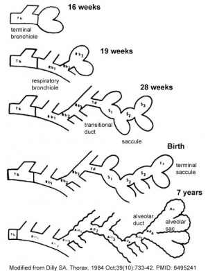
The sequence is most important rather than the actual timing, which is variable in the existing literature.
- week 4 - 5 embryonic
- week 5 - 17 pseudoglandular
- week 16 - 25 canalicular
- week 24 - 40 terminal sac
- late fetal - 8 years alveolar
Canalicular stage
- week 16 - 24
- Lung morphology changes dramatically
- differentiation of the pulmonary epithelium results in the formation of the future air-blood tissue barrier.
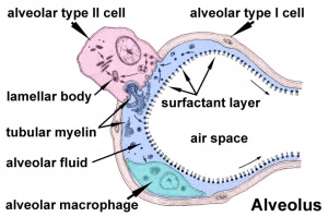
- Surfactant synthesis and the canalization of the lung parenchyma by capillaries begin.
- future gas exchange regions can be distinguished from the future conducting airways of the lungs.
Saccular stage
- week 24 to near term.
- most peripheral airways form widened airspaces, termed saccules.
- saccules widen and lengthen the airspace (by the addition of new generations).
- future gas exchange region expands significantly.
- Fibroblastic cells also undergo differentiation, they produce extracellular matrix, collagen, and elastin. May have a role in epithelial differentiation and control of surfactant secretion
- The vascular tree also grows in length and diameter during this time.
- in late fetal development respiratory motions and amniotic fluid are thought to have a role in lung maturation.
- Development of this system is not completed until late fetal just before birth.
- Therefore premature babies have difficulties associated with insufficient surfactant (end month 6 alveolar cells type 2 appear and begin to secrete surfactant).
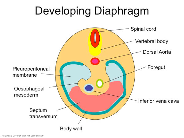
Diaphragm Development
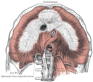
Five elements contribute to the diaphragm.
|
|
- Links: DiaphragmDevelopment
| 2011 Lab 5: Introduction | Trilaminar Embryo | Early Embryo | Late Embryo | Fetal | Postnatal | Abnormalities | Quiz | Online Assessment |
Glossary Links
- Glossary: A | B | C | D | E | F | G | H | I | J | K | L | M | N | O | P | Q | R | S | T | U | V | W | X | Y | Z | Numbers | Symbols | Term Link
Cite this page: Hill, M.A. (2026, February 27) Embryology 2011 Lab 5 - Fetal. Retrieved from https://embryology.med.unsw.edu.au/embryology/index.php/2011_Lab_5_-_Fetal
- © Dr Mark Hill 2026, UNSW Embryology ISBN: 978 0 7334 2609 4 - UNSW CRICOS Provider Code No. 00098G
