2010 Lecture 14
Limb Development
Introduction
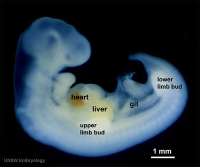
This lecture is an introduction to the events in limb development. Initially somites develop and then begin to differentiate forming sclerotome, dermomyotome and then dermatome and myotome. The lateral portion of the hypaxial myotome edge migrates at level of limbs (upper limb first then lower) and mixes with somatic mesoderm. Meanwhile the dermotome continues to contribute cells to myotome.
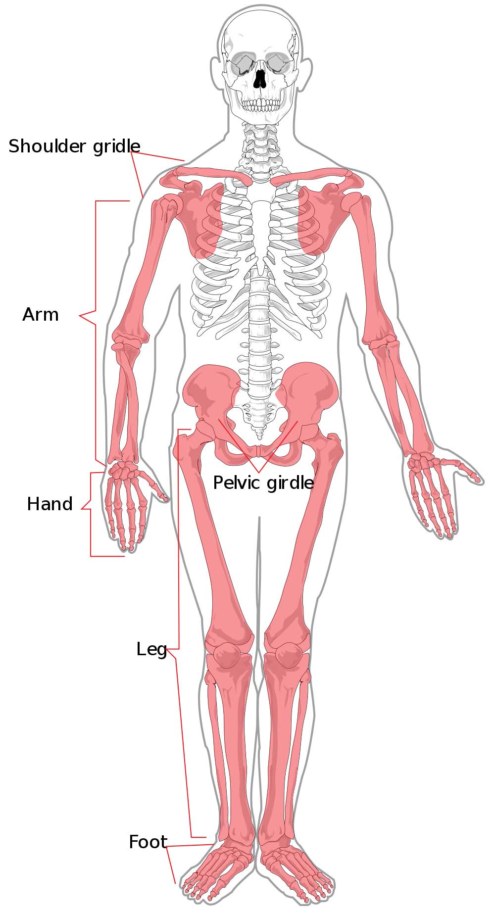
The appendicular skeleton consists of: Shoulder girdle, Upper limb (arm, hand), Pelvic girdle, Lower limb (leg, foot).
UNSW Embryology Limb Development | Limb Abnormalities 2008 Lecture 2008 Lecture | 1 slide/page | 4 slide/page | 6 slide/page
- Lectopia Audio Lecture Date: 15-09-2009 Lecture Time: 12:00 Venue: BioMed E Speaker: Mark Hill Limb
Lecture Objectives
- Understanding of limb positioning
- Understanding of differences in developmental timing of upper and lower limbs
- Understanding of regions and factors determining limb axes
- Understanding of limb rotation
- Understanding of limb muscle, blood vessel, bone and nerve formation
- Brief understanding of limb molecular factors and cell death
- Brief understanding of limb abnormalities
Textbook References
- The Developing Human: Clinically Oriented Embryology (8th Edition) by Keith L. Moore and T.V.N Persaud - Moore & Persaud Chapter 15 the skeletal system
- Larsen’s Human Embryology by GC. Schoenwolf, SB. Bleyl, PR. Brauer and PH. Francis-West - Chapter 11 Limb Development
- Before we Are Born (5th ed.) Moore and Persaud Ch16,17: p379-397, 399-405
- Essentials of Human Embryology Larson Ch11 p207-228
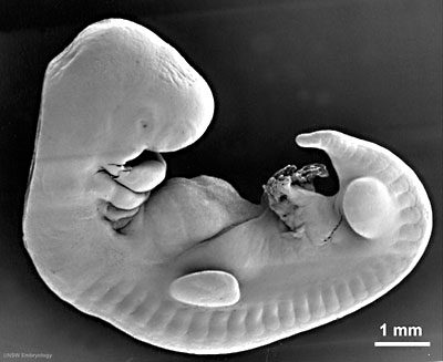
Limb Buds
- Limbs are initially undifferentiated mesenchyme (mesoderm) with an epithelial (ectoderm) covering.
- Blood vessels then begin forming, the largest (marginal vein) is adjacent to tip of the limbbud.
- Positioning of the limbs is distant from final location
Upper and Lower Limb
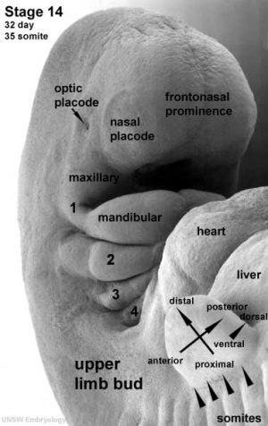
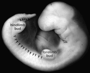
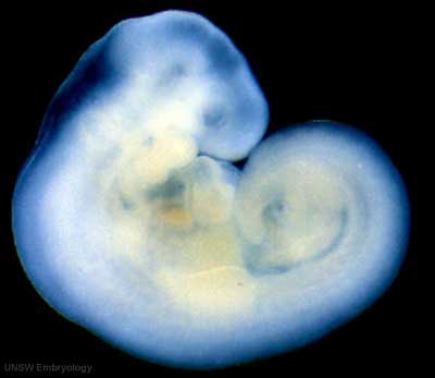
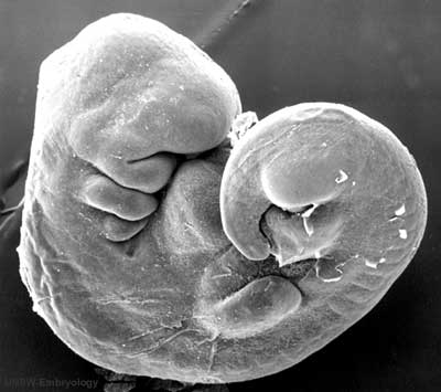
Limb development occurs at different times for forelimbs and hindlimbs. In the mid-4th week, human upper limb buds first form and lower limbs about 2 days later. The limbs form at vertebra segmental levels C5-C8 (upper limbs) L3-L5 (lower limbs).
Limb Axis Formation
Four Concepts - much of the work has been carried out using the chicken and more recently the mouse model of development.
- Limb Initiation
- Proximodistal Axis
- Dorsoventral Axis
- Anteroposterior Axis
Limb Initiation
- Fibroblast growth factor (FGF) coated beads can induce additional limb
- FGF10 is expressed in lateral plate mesoderm prior to bud formation induces expression of FGF8 in the overlying ectoderm. FGF8 induces continued growth in the underlying mesoderm - thus a positive feedback loop
- Anterior boundary of Hoxc6 expression coincides with the position of forelimb development
Autoregulatory loop of induction between FGF10 and FGF8
Site of FGF10 expression in the chick embryo
Examples of Hox gene expression boundaries in the mouse Hoxb2 and Hoxb4
Limb Identity
Forelimb and hindlimb (mouse) identity appears to be regulated by T-box (Tbx) genes, which are a family of transcription factors.
- hindlimb Tbx4 is expressed.
- forelimb Tbx5 is expressed.
- Tbx2 and Tbx3 are expressed in both limbs.
Related Research - PMID: 12490567 | Development 2003 Figures | Scanning electron micrographs of E9 Limb bud wild-type and Tbx5del/del A model for early stages of limb bud growth | PMID: 12736217 | Development 2003 Figures
Tbx4 expression can turn an experimentally induced forelimb into a hindlimb
Axes and Morphogens
- Anteroposterior - (Rostrocaudal, Craniocaudal, Cephalocaudal) from the head end to opposite end of body or tail.
- Dorsoventral - from the spinal column (back) to belly (front).
- Proximodistal - from the tip of an appendage (distal) to where it joins the body (proximal).
Model of patterning signals in the vertebrate limb
Diffusible morphogens create a concentration gradient accross an embryonic field
Proximodistal Axis
- Apical Ectodermal Ridge (AER) initially formed at the site of FGF10 induction
- then AER secretes FGF8 and FGF4 slightly later
- FGFs stimulate proliferation and outgrowth in the underlying mesenchyme
apical ectodermal ridge | AER and vascular channel
Fibroblast growth factors (FGF)
- 22 FGF genes identified in humans
- bind membrane tyrosine kinase receptors
- Patterning switch with many different roles in different tissues
FGF receptors
- comprise a family of at least 4 related but individually distinct tyrosine kinase receptors (FGFR1- 4) similar protein structure
- 3 immunoglobulin-like domains in extracellular region
- single membrane spanning segment
- cytoplasmic tyrosine kinase domain
FGF receptors are paired proteins on the cell surface with an internal tyrosine kinase domain
Dorsoventral Axis
- Ventral muscles - flexors Dorsal muscles - extensors
- Early grafting experiments showed that the D/V signalling centre resided in the dorsal ectoderm
- Wnt7a is a diffusible morphogen that is secreted by dorsal ectoderm cells
- Wnt7a induces the expression of the homeobox gene Lmx1 in the underlying mesoderm adjacent to the dorsal surface
- The homeobox gene Engrailed (En1) is expressed in the opposite ventral ectoderm
Consequence of Wnt7a deficiency in the mouse forelimb
Wnt7a
- name was derived from 'wingless' and 'int’
- Wnt gene first defined as a protooncogene, int1
- Humans have at least 4 Wnt genes
- Wnt7a gene is at 3p25 encoding a 349aa secreted glycoprotein
- patterning switch with different roles in different tissues
- One WNT receptor is called Frizzled (FZD) - named after a drosophila phenotype
- Frizzled gene family encodes a 7 transmembrane receptor
Anteroposterior Axis
- Zone of polarizing activity (ZPA)
- a mesenchymal posterior region of limb
- secretes sonic hedgehog (SHH)
ZPA secretes SHH and determines the anteroposterior axis of the limb bud
Dynamic development - time, the 4th dimension
- Different Hox genes are expressed at different times in the developing limb bud and pattern the fine structure of the limb.
- Structures are determined in a proximal>distal direction with time, i.e. proximal structures such as the humerus bone are laid down first.
Hox genes and dynamic patterning of the limb
Cellular origins of the limb
Limb cartilage and bone
- Derived from local proliferating mesenchyme derived from the somatic lateral plate mesoderm (somatopleure)
Limb muscle and dermis
- Skeletal muscle derived from somites, the hypaxial part of the myotome
- Pax3 positive migratory myoblasts invade the limb bud
- Similarly, dermal cells also invade derived from the dermomyotome
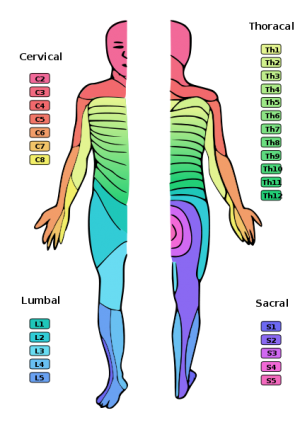
- Both maintain the identity of the somite from which they were derived so that innervation corresponds to the same spinal nerve root.
- Note that dermatomes are rotated due to embryonic limb rotations
Origin of limb muscle cells - Migrations traced by grafting cells from a quail embryo into a chick embryo
- two species very similar in development
- quail cells recognizable by distinctive nucleoli
- Quail somite cells substituted for somite cells of 2 day chick embryo
- wing of chick sectioned a week later
- found muscle cells in chick wing derive from transplanted quail somites
Dorsal/Ventral Muscle Mass
Forelimb Muscles
Limb Muscle - Differentiation, Skeletal muscle differentiates the same
- Muscle precursor cells migrate to the muscle location
- Form beds of proliferating myoblasts
- Myoblasts fuse together to form myotubes
- Myotubes begin to express contractile proteins, form sarcomeres
- mature into myofibers, Innervation determines final muscle maturation
Dermomyotome MyoD
Hand and Footplates
- 5th week- hand and footplates appear at the ends of limb buds and ridges form digital rays
- Cells between the digital rays are removed by programmed cell death (apoptosis)
- 3-5 day difference between hand and foot development
Apoptosis
Cell Biology - Cell Death Lecture | Cell Biology - Apoptosis Lecture
Fluorescent staining of cells undergoing apoptosis in the limb
Limb Rotation
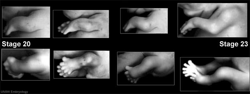
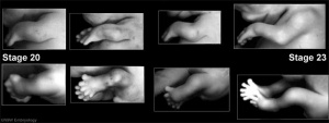
- 8th week limbs rotate in different directions (Humans Stage 20-23)
- thumb and toe rostral
- knee and elbow face outward
- upper limb rotates dorsally
- lower limb rotates ventrally
Limb Innervation
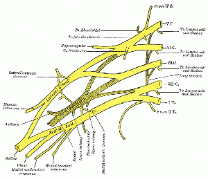
- spinal cord segmental nerves form a plexus adjacent to each limb
- Brachial (upper) lumbar (lower)
- Plexus forms as nerves invade the limb bud mesechyme
- Fetal period - touch pads become visible on hands and feet
Limb Abnormalities
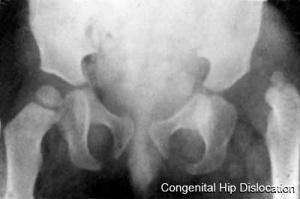
Congenital Hip Dislocation
- Instability: 1:60 at birth; 1:240 at 1 wk: Dislocation untreated; 1:700
- congenital instability of hip, later dislocates by muscle pulls or gravity
- familial predisposition female predominance
- Growth of femoral head, acetabulum and innominate bone are delayed until the femoral head fits firmly into the acetabulum
Maternal
- thalidomide Phocomelia
- short ill-formed upper or lower limbs
- hyperthermia
Genetic
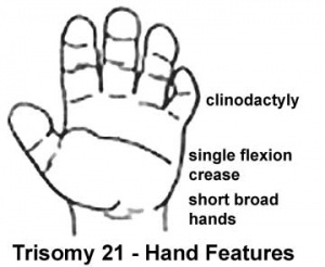
- Trisomy 21 - Downs syndrome
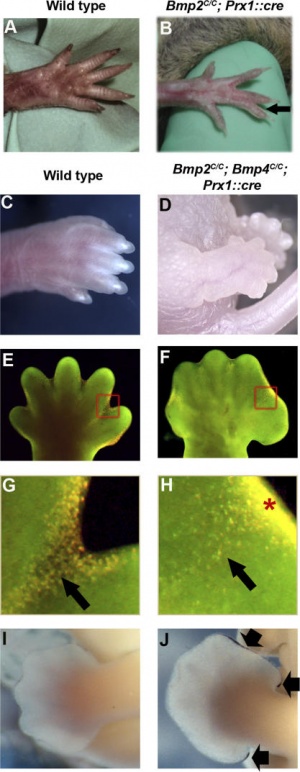
- Human Gene Mutations - mutation of any of the patterning genes will result in limb abnormalities
Type II syndactyly- HoxD13
Muscle Development
Duchenne Muscular Dystrophy
- X-linked dystrophy
- large gene encoding cytoskeletal protein- Dystrophin
- progressive wasting of muscle, die late teens
Becker Muscular Dystrophy
- milder form, adult onset
Online Links
- UNSW Embryology Limb Development
- Embryo Images Limb Unit
- International J. Dev. Biology Vol 46 Special Issue- Limb Development 2002
- Research Labs - Rolf Zeller University of Basel Medical School
References
Textbooks
- The Developing Human: Clinically Oriented Embryology (8th Edition) by Keith L. Moore and T.V.N Persaud - Moore & Persaud
- Larsen’s Human Embryology by GC. Schoenwolf, SB. Bleyl, PR. Brauer and PH. Francis-West -
Online Textbooks
- Developmental Biology by Gilbert, Scott F. Sunderland (MA): Sinauer Associates, Inc.; c2000 Formation of the Limb Bud | Generating the Proximal-Distal Axis of the Limb
- Molecular Biology of the Cell Alberts, Bruce; Johnson, Alexander; Lewis, Julian; Raff, Martin; Roberts, Keith; Walter, Peter New York and London: Garland Science; c2002 Figure 21-13. Sonic hedgehog as a morphogen in chick limb development
- Madame Curie Bioscience Database Chapters taken from the Madame Curie Bioscience Database (formerly, Eurekah Bioscience Database)
Search
- Bookshelf limb development
- Pubmed limb development
Images
Stage13
Stage14
Glossary Links
- Glossary: A | B | C | D | E | F | G | H | I | J | K | L | M | N | O | P | Q | R | S | T | U | V | W | X | Y | Z | Numbers | Symbols | Term Link
Course Content 2010
Embryology Introduction | Cell Division/Fertilization | Lab 1 | Week 1&2 Development | Week 3 Development | Lab 2 | Mesoderm Development | Ectoderm, Early Neural, Neural Crest | Lab 3 | Early Vascular Development | Placenta | Lab 4 | Endoderm, Early Gastrointestinal | Respiratory Development | Lab 5 | Head Development | Neural Crest Development | Lab 6 | Musculoskeletal Development | Limb Development | Lab 7 | Kidney | Genital | Lab 8 | Sensory | Stem Cells | Stem Cells | Endocrine | Lab 10 | Late Vascular Development | Integumentary | Lab 11 | Birth, Postnatal | Revision | Lab 12 | Lecture Audio | Course Timetable
Cite this page: Hill, M.A. (2026, February 28) Embryology 2010 Lecture 14. Retrieved from https://embryology.med.unsw.edu.au/embryology/index.php/2010_Lecture_14
- © Dr Mark Hill 2026, UNSW Embryology ISBN: 978 0 7334 2609 4 - UNSW CRICOS Provider Code No. 00098G
