2010 Lecture 10
Introduction


The respiratory system does not carry out its physiological function (of gas exchange) until after birth. The respiratory tract, diaphragm and lungs do form early in embryonic development.
The respiratory tract is divided anatomically into 2 main parts:
- upper respiratory tract - consisting of the nose, nasal cavity and the pharynx.
- lower respiratory tract - consisting of the larynx, trachea, bronchi and the lungs.
The respiratory "system" usually includes descriptions of not only the functional development of the lungs, but also related musculoskeletal (diaphragm) and vascular (pulmonary) development.
Links: Respiratory System Development
Lecture Objectives


- Understanding of embryonic lung development
- Understanding of 4 stages of lung development
- Understanding of diaphragm development
- Brief understanding of respiratory vascular development
- Brief understanding of respiratory abnormalities
- Brief understanding of molecular mechanisms
Textbook References
- Human Embryology Larson Chapter 9 p229-260
- The Developing Human: Clinically Oriented Embryology (6th ed.) Moore and Persaud Chapter 12 p271-302
- Respiratory System Development
Additional Textbooks
- Before We Are Born (5th ed.) Moore and Persaud Chapter 13 p255-287
- Essentials of Human Embryology Larson Chapter 9 p123-146
- Human Embryology Fitzgerald and Fitzgerald Chapter 19,20 p119-123
- Anatomy of the Human Body 1918 Henry Gray 1. The Respiratory Apparatus
- Developmental Biology 8e OnlineLung Branching Morphogenesis
Developmental Overview
Week 4 - laryngotracheal groove forms on floor foregut.
Week 5 - left and right lung buds push into the pericardioperitoneal canals (primordia of pleural cavity)
Week 6 - descent of heart and lungs into thorax. Pleuroperitoneal foramen closes.
Week 7 - enlargement of liver stops descent of heart and lungs.
Month 3-6 - lungs appear glandular, end month 6 alveolar cells type 2 appear and begin to secrete surfactant.
Month 7 - respiratory bronchioles proliferate and end in alveolar ducts and sacs.
Lung Development
- week 4 - 5 embryonic
- week 5 - 17 pseudoglandular
- week 16 - 25 canalicular
- week 24 - 40 terminal sac
- late fetal - 8 years alveolar
Germ Layers
- Endoderm and splanchnic mesoderm form majority of conducting and alveoli.
- Ectoderm will contribute the neural innervation.
- Mesoderm also contributes the supporting musculoskeletal components.
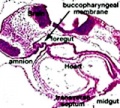
Foregut development
From the oral cavity the next portion of the foregut is initially a single gastrointestinal (oesophagus) and respiratory (trachea) common tube, the pharynx which lies behind the heart. Note that the respiratory tract will form from a ventral bud arising at this level.
- Oral cavity
- Pharynx (esophagus, trachea)
- Respiratory tract
- Stomach
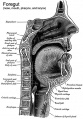
Upper respiratory tract

stage 11 foregut

week 4 early respiratory endodermal bud
Stage 22 trachea
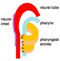
Head arches cartoon
Pharynx
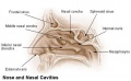
Nasal cavities
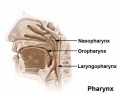
Pharynx
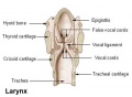
Larynx


- part of foregut development
- anatomically the nose, nasal cavity and the pharynx
- the pharynx forms a major arched cavity within the pharyngeal arches
MH - pharyngeal arches will be described in head development lecture
Lower respiratory tract
week 4 early respiratory endodermal bud

week 4 later ventral endoderm growth
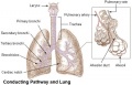
lower respiratory tract
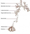
conducting system bronchi to lungs


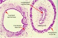
- lung buds ( endoderm epithelial tubes) grow/push into mesenchyme covered with pleural cells (lung border)
- generates a tree-like network by repeated:
- elongation
- terminal bifurcation
- lateral budding
- The lungs go through an embryonic and 4 distinct histological phases of development
Growth initially of branched "conducting" system of bronchial tree, followed by later development of the "functional units" of the alveoli.
- embryonic - week 4 - 5 (stage 14 above)
- pseudoglandular - week 5 - 17 (stage 22 above)
- canalicular - week 16 - 25
- terminal sac - week 24 - 40
- alveolar - late fetal - 8 years (Latin, alveus = cavity or hollow)
Fetal lung volume
Each lung volume as determined by ultrasound and matched to gestational age (PMID: 16388511)
- 12 - 13 weeks 0.05 mL
- 19 - 22 weeks 0.5 mL
- 29 - 32 weeks 1.9 mL
Pleural Cavity


- The anatomical body cavity in which the lungs develop and lie.
- The pleural cavity forms in the lateral plate mesoderm as part of the early single intraembryonic coelom.
- This cavity is initially continuous with pericardial and peritoneal cavities and form initially as two narrow canals
- later becomes separated by folding (pleuropericardial fold, pleuroperitoneal membrane) and the later formation of the diaphragm
pleuropericardial fold - (pleuropericardial membrane) An early embryonic fold which restricts the communication between pleural cavity and pericardiac cavity, contains both the cardinal vein and phrenic nerve.
pleuroperitoneal membrane - An early embryonic membrane that forms inferiorly at the septum transversum to separate peritoneal cavity from pleural cavity.
Pleura
- serous membrane covers the surface of the lung and the spaces between the lobes
- arranged as a closed invaginated sac
- two layers (pulmonary, parietal) continuous with each other, the potential space between them is the pleural cavity
Diaphragm
Not respiratory tract but musculoskeletal development, there are 5 elements that contribute to the diaphragm.

|
|


Innervation of the human diaphragm is by the phrenic nerves, arising from the same segmental levels from which the diaphragm skeletal muscles arise, segmental levels C3 to C5.
The paired phrenic nerves are mixed containing motor neurons for the diaphragm and sensory nerves for other abdominal structures (mediastinum, pleura, liver, gall bladder).
Pulmonary Circulation

- the pulmonary system not "functional" until after birth
- pulmonary arteries - 6th aortic arch arteries
- pulmonary veins - are incorporated into the left atrium wall
- bronchial arteries - branches from dorsal aorta
Fetal
Fetal Respiratory Movements
- Fetal respiratory movements (FRM) or Fetal breathing movements (FBM) are regular muscular contrations occurring in the third trimester.
- thought to be preparing the respiratory muscular system for neonatal function
- thought to also have a role in late lung development.
The First Breath

- The respiratory system does not carry out its physiological function (gas exchange) prenatally and remain entirely fluid-filled until birth.
- At birth, fluid in the upper respiratory tract is expired and fluid in the lung aveoli is rapidly absorbed this event has also been called "dewatering of the lung".
- The lung epithelia has to now rapidly change from its prenatal secretory function to that of fluid absorbtion.
The exchange of lung fluid for air leads to:
- fall in pulmonary vascular resistance
- increase in pulmonary blood flow
- thinning of pulmonary arteries (stretching as lungs increase in size)
- blood fills the alveolar capillaries
In the heart, pressure in the right side of the heart decreases and pressure in the left side of the heart increases (more blood returning from pulmonary).

- Respiratory Rate is higher than adult (30 breaths/minute).
- Rib Orientation - Infant rib is virtually horizontal, allowing diaphragmatic breathing only. Adult rib orientation is oblique (both anterior and lateral views), allows for pump-handle and bucket handle types of inspiration.
Respiratory Tract Abnormalities
Respiratory System - Abnormalities
Tracheoesophageal Fistula
(Tracheo-Oesophageal Fistula, Oesophageal Atresia) - Oesophageal Atresia with or without tracheo-oesophageal fistula
Lobar Emphysema (Overinflated Lung)
- There is an overinflated left upper lobe
- There is a collapsed lower lobe
- The left lung is herniating across the mediastinum
Congenital Diaphragmatic Hernia
Failure of the pleuroperitoneal foramen (foramen of Bochdalek) to close allows viscera into thorax. Intestine, stomach or spleen can enter the pleural cavity, compressing the lung.
Azygos Lobe

- Common condition (0.5% of population).
- The right lung upper lobe expands either side of the posterior cardinal.
- There is also some course variability of the phrenic nerve in the presence of an azygos lobe.
Congenital Laryngeal Webs
Laryngeal abnormality due to embryonic (week 10) incomplete recanalization of the laryngotracheal tube during the fetal period. Rare abnormality occuring mainly at the level of the vocal folds (glottis).
Meconium Aspiration Syndrome
(MAS) Meconium is the gastrointestinal contents that accumulate in the intestines during the fetal period. Fetal stress in the third trimester, prior to/at/ or during parturition can lead to premature meconium discharge into the amniotic fluid and sunsequent ingestion by the fetus and damage to respiratory function. Damage to placental vessels meconium myonecrosis may also occur.
Newborn Respiratory Distress Syndrome
(Hyaline Membrane Disease) medline plus | eMedicine
Bronchopulmonary Dysplasia
A chronic lung disease which can occur following premature birth. The definition of bronchopulmonary dysplasia (BPD) has in recent years changed from a severe lung injury and associated repair, to more of a disruption of lung development.
Glossary Links
- Glossary: A | B | C | D | E | F | G | H | I | J | K | L | M | N | O | P | Q | R | S | T | U | V | W | X | Y | Z | Numbers | Symbols | Term Link
Course Content 2010
Embryology Introduction | Cell Division/Fertilization | Lab 1 | Week 1&2 Development | Week 3 Development | Lab 2 | Mesoderm Development | Ectoderm, Early Neural, Neural Crest | Lab 3 | Early Vascular Development | Placenta | Lab 4 | Endoderm, Early Gastrointestinal | Respiratory Development | Lab 5 | Head Development | Neural Crest Development | Lab 6 | Musculoskeletal Development | Limb Development | Lab 7 | Kidney | Genital | Lab 8 | Sensory | Stem Cells | Stem Cells | Endocrine | Lab 10 | Late Vascular Development | Integumentary | Lab 11 | Birth, Postnatal | Revision | Lab 12 | Lecture Audio | Course Timetable
Cite this page: Hill, M.A. (2026, July 11) Embryology 2010 Lecture 10. Retrieved from https://embryology.med.unsw.edu.au/embryology/index.php/2010_Lecture_10
- © Dr Mark Hill 2026, UNSW Embryology ISBN: 978 0 7334 2609 4 - UNSW CRICOS Provider Code No. 00098G