1897 Human Embryology - Figures
| Embryology - 26 Jul 2026 |
|---|
| Google Translate - select your language from the list shown below (this will open a new external page) |
|
العربية | català | 中文 | 中國傳統的 | français | Deutsche | עִברִית | हिंदी | bahasa Indonesia | italiano | 日本語 | 한국어 | မြန်မာ | Pilipino | Polskie | português | ਪੰਜਾਬੀ ਦੇ | Română | русский | Español | Swahili | Svensk | ไทย | Türkçe | اردو | ייִדיש | Tiếng Việt These external translations are automated and may not be accurate. (More? About Translations) |
Minot CS. Human Embryology. (1897) London: The Macmillan Company.
- Note - this online text is only at a very early draft stage and contains many errors from the original scanning.
| Historic Disclaimer - information about historic embryology pages |
|---|
 |
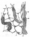
Fig. 1. Connective tissue of mucosa, uterus of pig
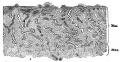
Fig. 2. Vertical section of the mucosa carpus uteri of the first day of menstruation
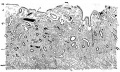
Fig. 3. Mucous membrane of a virgin uterus during the first day of menstruation
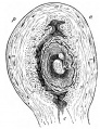
Fig. 4. Semi-diagrammatic outline of an antero-posterior section of the gravid uterus and ovum of five weeks
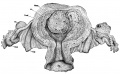
Fig. 5. Uterus about forty days advanced in pregnancy
Fig. 6. Uterus one month pregnant ; outlines of the glands from a vertical section
Fig. 7. Uterus one mouth pregnant ; portion of the compact layer of the decidua seen in vertical section
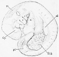
Fig. 216. His' embryo a, age probably twenty-three days
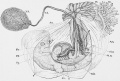
Fig. 222. Embryo of about thirty-five days
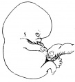
Fig. 224. Embryo of 22 mm
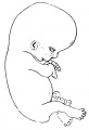
Fig. 225. Embryo of 28 mm.
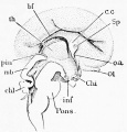
Fig. 391. Brain of a human embryo of about three months
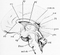
Fig. 392. Brain of a human embryo of the fourth month
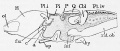
Fig. 393. Median view of a frog's brain
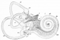
Fig. 429. Isolated right membranous labyrinth of human embryo of six months
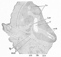
Fig. 430. Section through the region of the ear of a human embryo of three months
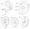
Fig. 431. Development of the human external ear
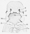
Fig. 432. Reconstruction of the pharyngeal region of a human embryo of 11.5 mm
Fig. 436. Reconstructions to show the development of the thyroid gland in the pig
Fig. 437. A section of the thyroid gland of a human embryo of about four months
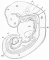
Fig. 441. Reconstruction of Fol's embryo
Fig. 444. Digestive tracts of four human embryos. A, embryo of 4.2 mm.; B, embryo of 7mm.; C, embryo of 13.8 mm.; D, embryo of 12.5 mm.
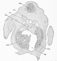
Fig. 451. Section of a rabbit embryo of thirteen days through the region of the fore limbs and liver
Fig. 463. Epithelium and gland of the trachea of a four months embryo


List of Illustrations
1. Connective tissue of mucosa, uterus of pig
2. Vertical section of the mucosa carpus uteri of the first day of menstruation
3. Mucous membrane of a virgin uterus during the first day of menstruation
4. Semi-diagrammatic outline of an antero-posterior section of the gravid uterus and ovum of five weeks
5. Uterus about forty days advanced in pregnancy
6. Uterus one month pregnant ; outlines of the glands from a vertical section
7. Uterus one mouth pregnant ; portion of the compact layer of the decidua seen in vertical section
8. Uterus one month pregnant ; section of gland froiu cavernous layer, with the epithelium partly adherent to the walls
9. Uterus one month pregnant ; section of gland from cavernous layer with the epithelium loosened from the walls
10. Section of the decidua serotina, near the margin of the placenta ; normal uterus about seven months pregnant
11. Decidual cells from the section represented in Fig. 10; stained with alum hsemotoxylin, and eosin
12. Section of human decidua reflexa at two months
13. Uterus twelve hours after artificial deUvery at six months' pregnancy
14. Section of the placental area of the uterus three weeks post partum
15. Vertical section through the wall of a uterus about seven months pregnant, with the foetal membranes in situ
16. Human embryo, 4.2 mm. long
17. Embryo, 2.15 mm. long
18. Diagram of an embryo of fifteen to sixteen days
19. Generalized diagram of an anmiote vertebrate embryo
20. Generalized diagram of an amniote vertebrate embryo before the separation of the amnion
2I. Structure of a rat's spermatozoon
22. Human spermatozoa
23. Peripheral layer of the seminiferous tubule of a rat
24. Column of spermatocytes from the rat
25. Developing spermatoblasts of the rat
26. Developing spermatozoa of a marsupial
27. Human spermatoblasts, to illustrate the rupture of the membrane
28. Sertoli's column, with a basal nucleolated nucleus and a cluster of developing si)ermatoblasts
29. Part of a cross-section of a seminiferous tubule of a rat
30. Egg of Tendra zostericola
31. Primary follicles from the ovary of a woman thirty-one years old
32. Ovary of cat
33. Egg-cell of Tengenaria domestica
34. Full-grown human ovum before maturation
35. Part of the ovum of a mole
36. Ovum of a sea urchin, Toxopneustes lividus
37. Ovarion egg of hsemops
38. Egg of a leech (nephelis), three-quarters of an hour after being laid
39. Ovum of nephelis (a leech), three hours after laying
40. Ovum of a rabbit ; taken from the middle of the oviduct about eighteen hours after coitus
41. Anterior pole of the ovum of the petromyzon, with a spermatozoon
42. Egg of nephelis, three hours after laying
43. Ovum of sagitta with two pronuclei
44. Two ova of the land-snail, arion
45. Ovum of a rabbit seventeen hours after coitus with the pronuclei about to conjugate
46. Ovum of Limax campestris during the first cleavage
47. Blastula stage of Echinocardium cordatum twenty hours after impregnation
48. Segmentation of the egg of the common frog
49. Section of the segmented ovum of axolotl
50. Four stages of the segmentation of the hen^s ovum
51. Ovum of a flounder in transverse vertical section
52. Ovum of a rabbit of twenty-four hours
53. Rabbit's ovum of about seventy hours
54. Ovum of a bat, Vespertilis murina, with four segmentation spheres
55. Ovum of Virginian opossum, with four segments
56. Young blastodermic vesicle of a mole
57. Sections through the inner mass of the blastodermic vesicle of the mole, at three successive stages
58. Ovum of a rabbit, ninety-four hours after coitus
59. Diagram of a seguiented mammalian ovum
60. Ovuui of Amphioxus lanceolatus during segmentation stage, with eighty-eight cells
61. Section of a erastrula of Toxopneustes lividus
62. Diagrams of the principal modiflcations of the gastrula
63. Longitudinal section of an early stage of the gecko
64. Diagram illustrating the growth of the blastoderm and concrescence of its rim to form the primitive axis
65. Diagram of concrescence in a teleostean egg
66. Diagram of an elasmobranch blastoderm to illustrate the formation of the marginal groove
67. Diagram of a vertebrate blastodenu a little more advanced than Fig. 96
68. Ovum of axolotl
69. Ovum of i>etromyzon in longitudinal section
70. Longitudinal section of the ovum of a sturgeon after the formation of the entodermic Cavity
71. Formation of the blastoporic canal in Lacerta mural is
72. Hen's o^1lm ; incubated six hours
73. Diagrammatic cross-section of a vertebrate ovum, in which concrescence is supposed to have been arrested
74. Dog-fish embryo, nearly in Balfour's stage C
75. Germinal area of a guinea-pig at thirteen days and twenty hours
76. Diagram showing the relations of a vertebrate ovum with an embryo in cross-section and a large yolk
77. Sections of axolotl eggs
78. Area pellucida of a hen's egg, with completed primitive furrow
79. Longitudinal section of the region of the primitive streak of a hen’s ovum incubated six hours
80. Transverse sections of a germinative area, with half-formed primitive streak, of a hen's egg
81. Transverse section of the anterior region of a fully developed primitive streak of a hen's ovum
82. Blastodermic vesicle of a rabbit of seven days
83. Transverse section of the embryonic shield of the blastodermic vesicle of a sheep
84. Central portion of a sheep's blastoilermic vesicle of twelve to thirteen days
85. Embryonic shield of a rabbit's ovum of five days
86. Section of the primitive streak of the mole
87. Blast<H]ermic vesicle of Mus sylvaticus
88. Axolotl embryo ; transverse section of an early stage
89. Diagrams of the embryonic area of the chick
90. Diagram of the embryonic area of a chick
91. The mesdermal cavities of the germinal area of a chick of the third day
92. Section of a chicken embryo of about thirty-six hours
93. Transverse section of an amphioxus embryo
94. Amphioxus embryo
95. Opossum embryo of seventy-three hours ; transverse section at the level of the heart
96. Blastoderm of rabbit's ovum
97. Chicken embryo with seven primitive segments
98. Part of a transverse section of a young mole embryo
99. Surface view of a young mole embryo
100. Transverse section of a mole embryo
101. Elarly stage of Amblystoma punctatum
102. Part of a transverse section of an axolotl embryo
103. Transverse section of a rabbit embryo of eight days and two hours
104. Part of a transverse section of an embryo of Lumbricus trapezoides
105. Transverse section of a mole embryo
106. Longitudinal section of the head end of a mole embryo
107. RaVibit embryo of 6 mm.; median longitudinal section of the head
108. Longitudinal sections of the notochord of bombinator
109. DegeneraHng notochord tissue, from the central portion of the intervertebral disc of a cow's embryo
110. Longitudinal section of a frog's ovum, shortly after closure of the medullary groove
111. Transverse section of an embryo paroquet (melopsittacus) to show the anterior or true ueurenteric canal
112. Chicken embryo with one segment
118. Area vasoalosa and embryo with eight segments of a hen's egg
114. Rabbit embryo with eight segments
115. Transverse section of a pristiurus embryo with fourteen segments, through the centre of the fourth segment
116. Transverse section through a recently formed primitive segment of a chicle with eight.een and twenty segments
117. Section of a chick with about twenty segments
118. Head of an embryo of Torpedo ocellata, in Balfour's stage J,
119. Longitudinal vertical section through five primitive segments of a rabbit embryo of nine days and seventeen hours
120. Longitudinal horizontal section through a segment of a rabbit embryo of ten and one-half days
121. Transverse section through the upper i>art of a myotome of a chick of about seventy hours
122. Pristiurus embryo with forty-flve to forty-six segments
123. Diagram of a cross-section of a young amphioxus
124. Surface view of a small part of the vascular network of an embryo chick of two days
125. Vascular anlages of the area vasculosa of a chick of forty hours
126. Section of the area vasculosa of a chick
127. Corpuscles from rabbits, from acanthias, from a chick, from a human embryo
128. Salamandra maculosa ; larva, very young ; transverse section to show the formation of the coelom in the heart region
129. Salamandra maculosa, larva with branchial arches,
129 A. Embryo chick ; section through the anlage of heart
130. Chick embryo
131. Diagrammatic cross-section of a vertebrate to show the fundamental relations of the urogenital system
132. Rana temporaria. Tadpole of 12 mm. Cross-section through the pronephros
133. Nephridium (or Wolffian tubule) of an acanthias embryo of 28.2 mm. , seen from the caudal side ; reconstructed from the sections
134. Section through a Wolffian tubule of a chick with primitive segments, 288
135. Wolffian tubule of a sheep embryo of 9 mm
136. Coste's embryo of thirty-five days
137. Transverse section of the Wolffian body or priiuitive kidney of a rabbit of thirteen days
138. Longitudinal vertical section of the AVolffian body of a rabbit embryo of thirteen days
139. Section through the testis of a human embryo of sixty-three to sixty eight days
140. Transverse section through an advanced embryo of a shark, sy<;m nus lichia ; from the abdominal region (dots represent nuclei)
141. Section of the urogenital fold of a chick embryo of the fourth day
142. Diagrammatic section of the yellow of a hen's egg at an early stage to show the relations of the arehenteron to the yolk-sac
143. Diagrams to illustrate the separation of the embryo from the yolk
144. Cross-section of a rabbit embryo of eight days and two hours
145. Lon^tudin&l seotion of the posterior end of a sheep embryo of sixteen days
146. Longitudinal median section of young chick embryo
147. Transverse section of the head of a chick embryo with seven segments
148. Two views of a wax model of the cavity of the pharynx of a rabbit embryo of eleven days, 264
140. Acanthias embryo of 17 mm. Horizontal section of the anterior half
150. Chicken embryo of sixty-eight hours
151. Acanthias of 17 mm.
152. Cross-section of a branchial arch of an advanced shark embryo
153. Longitudinal section of an embryo of Petromyzon planeri, four days old, reared at Naples
154. Diagrams to indicate the fundamental relatione of the archenteron
155. Chicken embryoand germ area after twenty-seven hours incubation
156. Embryonic area of a rabbit of eleven days, with the placental area partly torn off
157. Diagram of the circulation in a chick at the end of the third day, as seen from the under or ventral side
158. Area vasculosa and embryo of a rabbit
159. Transverse section of the rump of a dog-fish embryo 14 mm. long
160. Section through the rump of a rabbit embryo of eight days and three hours, 282
161. Transverse section of the rump of an embryo chick of the third day, 283
162. Diagrams to illustrate His' theory of the origin of the human amnion
163. Reichert's ovum. Two views engraved from the original plate
164. Cross-section of Spec's embryo
165. Section passing through the blastopore of Spec's embryo
166. Diagram of His' embryo E : age fourteen (?) days; length about 2.3 mm
167. Thomson's second ovum
16lS. Human embryo of thirteen to fourteen days
169. Embryo of the beginning of third week
170. Human embryo of 2.15 mm. ; anatomy reconstructed from the sections
171. His' embryo L, 2.4 mm. long
172. Ovum supposed to be from fifteen to eighteen days old
173. Embryo supposed to be from fifteen to eighteen days old
174. Fragment of the chorion of fig. 4, highly magnified
175. His' embryo M
176. Digestive canal of His' embryo
177. Anterior wall of the pharynx of His' embryo BB, 3.2 mm. long
178. W. His' embryo M
179. Reconstruction of His' embryo BB, 3.2 mm. long
180. Reconstruction of His' embryo
181. Isolated tenninal branch of a villus from the chorion of an embryo of twelve weeks
182. Villous stem from a placenta of the fifth month
183. Terminal villi of a placenta at full term
184. Section of the chorion at three weeks
185. Aborting villus from a chorion of the second month
186. Placental chorion of an embryo of seven months
187. Section of the chorionic membrane of an ovum supposed to belong to the third week
188. Section of the chorionic membrane of an embryo of three weeks
189. Section of the amnion and placental chorion of the fifth month
190. Adenoid tissue of a villus from a placenta of four months
191. Section of the amnion covering the placenta of a two months embryo
192. Two sections of the placental amnion
193. A natural group of nuclei from the mesoderm of the amnion of a fetus of the fifth month
194. Mesodermic nuclei of the amnion of an embryo of about four months
195. Surface view of the amniotic epithelium of an embryo of 144 days
196. Diagram of the development of the foetal adnexa in the rabbit
197. Longitudinal median section of a petromyzon larva
198. Wall of the yolk-sac in the area opaca of a chick of the second day
199. Section of the yolk-sac of a human embryo
200. Diagram of the embryo and yolk-sac of a rabbit
201. Vertical section of the wall of the yolk-sac of a rabbit embryo of thirteen days
202. Diagram of an opossum embryo and its appendages
203. Section of the allantois from the umbilical cord of an embryo of three months
204. Diagrammatic section of the bauclisliel of a human embryo, modified from W. His
205. Sections of human umbilical cords
206. Connective tissue of the umbilical cord of an embryo of 21 mm
207. Connective tissue of the umbilical cord of a human embryo of about three months
208. Epithelial covering of the umbilical cord of an embryo of three months
209. Cross-section of an umbilical cord at term
210. Placenta at full term, doubly injected by Dr. H. P. Quincy to show the distribution of the vessels upon the surface
211. Placenta at full term
212. Mesenchymal tissue of a villus, from a placenta of four months
213. Section through a normal x)lacenta of about seven months, in situ
214. Portion of an injecte<l villus from a placenta of about five months
215. Placenta of about i\\e months ; portion of a small villus
216. His' embryo a, age probably twenty-three days
217. FoFs embryo of 5.6 mm., probably twenty-five days old
218. His' Embryo A, 7.5 mm. longm
219. Embryo of 9.8 mm.m
220. Embryo of about 14 mm
221. Dorsal view of an embryo of about 14 mm.
222. Embryo of about thirty-five days
223. His' embryo XXXI V,
224. Embryo of 22 mm
225. Embryo of 28 mm
226. Embryo of 32 mm
227. Embryo of 34 mm
228. Embryo of 55 mm
229. Embryo of 78 mm.
230. Front view of the head and face of the embryo
381. Embryo of about 120 mm
282. Embryo of 118 mm
238. Embryo of 155 mm
234. Mesenchyma of a chick embryo of the third day from close to the otocyst
285. Omentum of a human embryo of five months
236. Parietal bone of a human embryo of fourteen weeks
237. Transverse section of the mandible of a human embryo of the tenth week
238. Prom a section of an ossifying vertebra of a human embryo of four months
239. Section of a vertebra of the same embryo at right angles to the plane of fig. 238, and corresponding in level to the lower part of the bracket L, fig. 238
240. Artery from the allaiitois of a chick, surrounded by a network of lymphatics
241. Section of the spleen of a human embryo of six months
242. Pat island from the skin of a human embryo of five months
243. Reconstruction of the last occipital, and first two cervical vertebra of a cow embryo of 8.8 mm.
244. Cross-section of the anlage of second cervical vertebra of a cow embryo of 8.8 mm
245. Longitudinal median section of the upper portion of the vertebral column of a cow embryo of 22.5 mm.
246. Prontal projection of the cephalic part of a vertebral column of a cow embryo
247. Embryo pig of about 16 mm
248. Embryo pig« one and one-third inch long
249. Section of the anterior portion of the snout of an embryo pig
250. Embryo pig, six inches long
251. Chondrocranium of an insectivorous mammal (Tatusia)
252. Pectoral fin of a young embryo of sycUium in longitudinal and horizontal section
253. Scapulaof a human embryo of five and one-half inches, dorsal view
254. Vertical section of the ankle of a human embryo of nearly six months
255. Isolated muscle fibres of a frog embryo
256. A, transverse section ; 6, longitudinal section of muscle fibres in the neck of a human embryo of sixty-three to sixty-eight days
257. Chick embryo, transverse section of the upper part of a myotome
258. Transverse section of a branchial arch of a selachian embryo
259. His^ embryo R, 5 mm. Reconstruction to show the septum transversum
260. Head of a rabbit embryo, with segments seen from the under side
261. Rabbit embryo, eight and a half days, with eleven or twelve somites, cross-section
262. Model of part of the pleural and abdominal cavities of a rat embryo at a stage corresponding to a rabbit at fifteen days
263. Section of the supra-renal body of a rabbit embryo of twenty-six days
264. Supra-renal capsule of a four months human embryo
265. Diagram of the indifferent stage of the urogenital system of amniota
266. Diagram to illustrate the homologies of the sexual apparatus
267. Section of the testis of a human embryo of sixty-three to sixty-eight days
268. Section of the ovary of a human embryo of 7 cm.
269. To illustrate the (lecensus te>ticulon]m
270. Cross-section of the ovarj' and Wolffian body of a human embryo of the third month
271. Cross-section of the rectum, genital cord, and allantois of a male human embryo of almost two months
272. Section of broad ligament of a female human embryo of four months
274. Crosft-section through the hind end of the left Wolffian body of a cr^jc/xlile embryoo of 12 mm
274. Section of a kidney, human embryo of about five months
275. Semidiagrammatic figures of developing renal tubules of a mammal
276. Section fiarallel to the medullary rays of the kidney of a human f^jftus of about five months,
277. Cross-section of the medullary tubules of the kidney of a human embryo of almost five months
278. Longitudinal median section of the cloaca of a sheep embryo of 18 mm
279. Longitudinal section of the penis of a human embryo of about five months
280. External genitalia, female embryo of 105 mm
281. Section of the clitoris and labia majora of a human embryo of about four and one-half months
282. Rxternul genitalia of the female human foetus at about four months
283. Head of chirk of thirty-eight hours, seen from the under side
284. Reconstruction of the heart and veinsof a human embryo of 2.15 mm.
284 A. Endothelial heart of a human embryo of 2.15 mm.; seen from the left side
285. Eleconstructed side view of the endothelial heart of a human embryo of 4.2 mm.
287. Model of the muscular heart of a rabbit embryo of nine to nine and one-half days, seen from the left side
288. Endothelial heart of a human embryo of 5 mm.
289. Inner surface of the heart of a human embrvo of 10 mm.
290. Section of the heart and pericardial cavity of a rabbit embryo of ten and one- half days
291. Section in the frontal plane through the heart of a rabbit embryo of thirteen days
292.Oblique section of the heart of a human embryo of 8.5 mm
293. Sections of different levels through the cardiac aorta of a human embryo of 11.5 mm
294. A diagram of pharynx of an amniote vertebrate
295. Anterior wall of the pharynx of a human embryoof 3.2 mm. length
296. Aortic HVHteni of His' embryo Bl., 4.25 mm
297. Aortic system of His' embryo Si, 12.5 mm.; seen from the front
298. Aortic system of W. His' embryo Rg, 11.5 mm
299. Reconstniction of the arteries of the head and neck of a rabbit embryo at the end of the eleventh day
300. His' embryo Lr (4.2 nnn.). Reconstruction to show the course of the blood-vessels
301. Cross-section through the hinder part of His' embryo R (5 mm.)
302. Three diagrams to illustrate the transformation of the venous system
303. lieconstruction of a human embryo (His' Bl.) of 4.25 mm
304. Reconstruction of the venous trunks and liver of His' embryo R, 5 mm
305. Reconstruction of the venous system of His' embryo Rg, 11.5 mm
306. Section of the skin of a human embryo of sixty-three to sixty-eight days
307. Epidennis from the occiput of the human embryo of two and one half months
308. Section of the skin of the under side of the right second toe of four months' embryo
309. Epitrichium of a human embryo of the fifth month
310. Vertical section of the skin of a human embryo of the fifth month
311. Longitudinal section of the nail of the great toe of a human embryo of five months
312. Development of hairs in a human embryo of about seven months
313. Isolated epidermis of a human embryo of five to six months
314. Section of the sole of the foot of a foetus of the fifth month, to show the sweat, glands
315. Development of the mammary gland in the rabbit
316. Acanthias embr\'oof 17 mm., under side
317. Blast odenii of a dog-fish, acanthias. with commencing tioncrescence
318. Longitudinal median section of a recently hatched larva of petromvzon
319. Longitudinal section of an acanthias embryo of 13.2 mm
320. Median section of the head of a rabbit embryo of thirteen and on half days
321. His' embryo A, 7.5 mm.
322. Fiicial region of a human embryo of 8 mm., front view
323. Reconstruction of the face of His' embryo Sell,
324. View of the roof of the mouth of a human embryo
325. Frontal section of the oral and nasal chambers of a young cow embryo
326. Frontal section of the nasal and oral cavities of a human embryo of three months
327. Dental papilla of a dermal tooth of an acanthias embryo of 10 cm
328. Section of the lower jaw of an acanthias embryo of 10 cm.
329. Section of part of the lower jaw of a human embryo of 40 mm.
330. Explanation in text
331. Vertical section of a molar tooth -germ of a human embryo of 160 mm.
332. Part of the enamel organ of a new-born child, incisor germ
333. Odontoblasts from cow embryos. A, of 30 cm.; B, of 24 cm.
334. Section of the submaxillary gland of a human embryo of sixtythree to sixty-eight days,
335. Reconstruction of the pharynx of a human embryo
336. Chick embryo of twenty-nine hours
337. Cross-section through the fore-brain and optic vesicles of a lepidosteus embryo of eight days
338. Brain of embryo No. 22, p. 297
339. Reconstruction of the brain of His' embryo Ko
340. Reconstructed median view of the fore-brain of His' embryo Ko
341. Brain of a human embryo of five weeks
342. Hind-brain of a human embryo
343. Dorsal view of the hind-brain of a human embryo of one month
344. Sections through the cervical part of the medulla of a human embryo with thirteen segments
845. Longitudinal horizontal section of the wall of the hind-brain of a young embryo of a lizard (Anolis Sagrsei)
346. Diagrammatic section of the embryonic spinal cord
347. Section of the medulla and otocysts
348. Sections through the regions 3 and 5 of the hind-brain of His^ embryo, 608
349. Sections through the region 3 of the hind-brain of His' embryo A, 609 850. Four sections of the brain of a human embryo of about five weeks, 609
351. Brain of His' embryo Br. 3, 610
352. Neuroglia of the dorsal zone of the spinal cord of a human embryo of about three and one-half weeks, 613
353. Cross-section of the spinal cord of a human embryo of 14 mm., to show the neuroglia cells
354. Part of a transverse section of the spinal cord of a human embryo of 23 cm
355. From a section of the medulla oblongata of His' embryo Br*
356. Group of motor neuroblasts and nerve fibres from a transverse section of the spinal cord of a cat embryo of ?? mm
357. Bipolar cells from a spinal ganglion of an embryo
358. Transverse section of the dorsal cord and ganglion of a chick of nine days
359. Isolated nerve fibres from the sciatic nerve of a sheep embryo of 150 mm
360. Part of the nerves of a human embryo of 13.8 mm
361. Cells and nuclei from the cervical region of the spinal cord of a human embrvo of one hundred and sixty days
362. Spinal ganglion cells from a longitudinal horizontal section of a human embryo of the tenth week
863. Peripheral nervous system of a human embryo of about 10 mm.
364. Transverse sect ir)n of a mouse embryo of about seventeen to eighteen days through the lumbar region
365. Transverse section of the sympathetic cord from the lower dorsal region of a rat embryo of about thirteen days
306. Sympathetic ganglia of one side of a human embryo of the fifth month
307. Transverse section through the posterior part of the mid-brain of a human embryo of five weeks
308. Section of the brain of a five weeks' embrvo
diV.i. Section of the brain of a five weeks' embryo
870. Section of the brain of a human embryo of five weeks
371. Otocvsis aii<l nerves of a human embrvo of four and one-half week
372 Accmstie ganglia of a human embryo of two months
373 Torpedo embryo of 12 mm
374. Section of the medulla oblongata of a five weeks' human embryo
375. Lower end of the spinal <'ord f»f a human embryo of three months
376. Section of the spinal cord of a human embryo of sixty-three to sixty-eight days no. Pjo
377. Transverse section of the spinal cord from the upper dorsal region of a human embryo of six weeks
3TS. Ix>wer cervical cord of a human embryo of about five months
379. Transverse section of the medulla oblongata of His' embryo Ru
380. Transverse section of the medulla oblongata of His' embryo Mr
381. Section through the medulla oblongata of His' embryo CR
382. Median section of the brain of a chick embryo of about four days
383. Longitudinal med^n section of the cerebellum of a chick of about twelve days
384. Section through the cerebellum and medulla oblongata of a human embryo of one hundred and sixty days
380. Section of the cerebellum of a human embryo of one hundred and sixty days
386. Median section of the head of a sheep embryo of .86 mm.
387. Brain, human foetus, five months
388. Part of the brain of His' embryo CR, 13.6 mm.
389. Section of the thalamencephalon of an embryo of five weeks
390. Section of the fore-brain of a sheep embryo of 27 mm.
391. Brain of a human embryo of about three months
392. Brain of a human embryo of the fourth month
393. Median view of a frog's brain
394. Section through the fore-brain of a foetal guinea-pig
395. Reconstruction of the brain of an embryo of about seven and onehalf weeks
390. Brain of a chick embryo, fourth day
397. Human embryo of about four months ; brain in situ
398. Section through the lateral wall of the cerebral hemisphere of a human embryo of four months
399. View of the hemisphere of a human embryo from the early part of the third month
400. Outlines of the fissure of Sylvius of human embryos at successive lunar months
401. Median view of the fore-brain of a human embryo from the beginning of the third month
402. Brain of human embryo of the fifth month after removal of the right hemisphere
403. Right hemisphere, natural size of a foetus of nearly seven months
404. Under side of the bniin of a human embryo of the fifth month
405. Section of the fore-brain of a human embryo of nearly five weeks
406. Horizontal section of the ciliary ganglion of a young torpedo embryo
407. Reconstruction to show the cephalic ganglia of a petromyzon larva 4 mm. long
408. Rabbit embryo of ten and one-half days ; section of head
409. Riibbit embryo of thirteen days; section of the eye
410. Reconstruction fnMii His' embryo Sch. 13.8 mm.
411. Section through the iris region of the eyelid a chick of thirteen days
412. Rabbit embryo of ten and one-half days : section of the lens anlage
415. Vertical section of the eye of a chick embryo of the third day
414. Section of the distal portion of the optic nerve of a rabbit embryo of thirteen days
415. Surface view of the membrana limitans externa with the developing rods and cones of a chick of fifteen to sixteen days
416. Injected vascular membrane of the retina of the eye of a pig embryo, 16 cm. long
417. Section through the iris region of the eye of a chick of thirteen days
418. United eyelids of a human embryo of about four months, seen in vertical section
419. Sections of human embryos showing the otocyst ; A, embryo of 2.4 mm.; B, embryo of 4 mm.
420. Horizontal section of the otocyst of a chick of the third day
421. Left otocyst of a human embryo of about four weeks ; A, from the inner, B, from the outer side
422. Transverse section of the head of a rabbit embryo of ten and one half days
423. Left otocyst of a human embryo of about five weeks, seen from out side and below
424. Transverse section of the semicircular canal of an embryo rabbit of twenty-four days
425. Left otocyst of a human embryo of about two months
426. Transverse section of scala media cochleee of a rabbit embryo of 55 mm.
427. Section through Corti's organ of the lower coil of the cochlea of a rabbit embryo of 75 mm.
428. Section through the internal ear of a sheep embryo, 28 mm.
429. Isolated right membranous labyrinth of human embryo of six months, seen from in front and outside
430. Section through the region of the ear of a human embryo of three months
431. Development of the human external ear ; A, embryo of one month ; B, six weeks ; C, eight weeks ; D, ten weeks ; E, fourteen weeks
432. Reconstruction of the pharyngeal region of a human embryo of 11.5 mm.
433. From a section of a tonsil of a human embryo of five months
434. Section through the third gill-cleft of a human embryo from the beginning of the third week
435. Reconstruction of the pharyngeal region of a human embryo of 9.1 mm.
430. Reconstructions to show the development of the thyroid gland in the pig; A, embryo of 15 mm.; B, of 16 mm.; C, of 20 mm.; D, of 22.5 mm
437. A, section of the thyroid gland of a human embryo of about four months ; B, a single acinus, more highly magnified
438. Reconstruction of His' embryo B ; the head is drawn as if erected
439. Transverse section of the oesophagus of a human embryo of four months
440. Highly magnified view of a small portion of the epithelium of fig. 4;^9,
441. Reconstruction of Fol's embryo
442. Epithelium of the greater curvature of the stomach of an embryo cat of 85 mm.
443 Peptic glands from the greater curvature of stomach of a human embryo from the end of the eighth lunar month
444. Digestive tracts of four human embryos. A, embryo of 4.2 mm.; B, embryo of 7mm.; C, embryo of 13.8 mm.; D, embryo of 12.5 mm.
445. Two front views of the entodenual canal. A, embryo Sch. 1 of His' B, His' embryo Sch. 2
440. Part of the inte8tine of a human embryo of about six months
447. Section of the small intestine of a human embryo of sixty-three to sixty -eight days
44S. Section of the small intestine of a human embryo of three months
440. Portion of a section of the liver of an acanthias embryo of 20 mm.
450. Section through the liver of a rabbit embryo of thirteen days
451. Section of a rabbit embryo of thirteen days through the region of the fore limbs and liver
452. Section of the t>ancreas of a human embryo of four months
453. Two diagrams to illustrate mori>ho]ogical relations of the vertebrate mesentery ; A, earlier ; B, later condition
454. Diagram to illustrate the relations of the mesentery
455. Diagram of the human mesentery in its primitive relations
456. Diagrams to illustrate the history of the human mesentery. A, earlier ; B, later condition
457. Two diagrams to illustrate the history of the mesentery; A, earlier ; B, later stages
458. Outline of the entodermal canal of His' embryo Lr.
460. Three views of the lungs of a human embryo of 10.5 mm., . 775 4<>0. Lungs of a human embryo of Hve months
461. Cross-section of the bronchial tube of a human embryo of sixty three to sixty-eight days
462. Section through the lung of a human embr>'o of the fourth month, 777
463. Epithelium and gland of the trachea of a four months embryo
| Historic Disclaimer - information about historic embryology pages |
|---|
|
Cite this page: Hill, M.A. (2026, July 26) Embryology 1897 Human Embryology - Figures. Retrieved from https://embryology.med.unsw.edu.au/embryology/index.php/1897_Human_Embryology_-_Figures
- © Dr Mark Hill 2026, UNSW Embryology ISBN: 978 0 7334 2609 4 - UNSW CRICOS Provider Code No. 00098G