User:Z3418837
Lab Attendance
- Lab 1 --Z3418837 (talk) 12:45, 6 August 2014 (EST)
- Lab 2 --Z3418837 (talk) 11:11, 13 August 2014 (EST)
- Lab 3 --Z3418837 (talk) 11:12, 20 August 2014 (EST)
- Lab 4 --Z3418837 (talk) 12:55, 27 August 2014 (EST)
- Lab 5
- Lab 6 --Z3418837 (talk) 12:44, 10 September 2014 (EST)
- Lab 7 --Z3418837 (talk) 12:40, 17 September 2014 (EST)
- Lab 8
- Lab 9
- Lab 10
- Lab 11
- Lab 12
Individual Assessment
Lab 1
--Z3418837 (talk) 12:45, 6 August 2014 (EST)
http://www.ncbi.nlm.nih.gov/pubmed
<pubmed>25084016</pubmed>
Your Lab assessment now requires you to find a 2 recent research references on fertilisation or in vitro fertilisation. Paste each reference on your page, as shown in the class. Write below each reference a brief summary of the research article methods and findings. The summary for each need not be more than 3-4 paragraphs in length. This will need to be completed before next weeks laboratory.
Reference:PMID25077107
<pubmed>25077107</pubmed>
This study was designed to investigate whether the levels of vitamin D is an imperative factor when it comes to the clinical success of implantation and pregnancy rates in infertile women via invitro fertilisation.
Method summary
A cohort of 173 women were evaluated and selected for this study based on their age, follicle-stimulating hormone levels and their consent to undergo invitro fertilisation. The following study was conducted at Mount Sinai Hospital where proper facilities were available. Blood tests were then conducted for each patient to determine their levels of Vitamin D via the serum 25-hydroxy-vitamin D (25[OH]D) levels. Following the results of the blood test, the cohort were then categorised into two groups which was either vitamin D sufficient (≥ 75 nmol/L) or insufficient ( < 75 nmol/L) based on serum levels of 25(OH)D.
Each patient then underwent IVF cycles whereby standard agonists that contained the active ingredient 0.5 mg/d of buserelin acetate in conjunction with cetrolix acetate as the standard antagonist were used to control the length of the luteal phase and estradiol levels. The length and dose of the treatment were varied for each individual based on their demographic data. Serial transvaginal ultrasonograpy and serum lutenizing hormone assays were then used to check ovarian response. When 3 or more dominant follicles (≥ 17 mm) were produced, 10 000 IU of human chorionic gonadotropin was added to enhance nuclear maturation. Oocyte retrieval was then conducted via transvaginal ultrasound whereby it was fertilised and the resulting embryo was transferred 3-5 days post-fertilisation. The rate of pregnancy per IVF cycle was then used as the primary outcome for this study whereby the visibility of the intrauterine sac of the embryo determined implantation.
Results summary
Out of the 182 patients that participated in this study, it was found that only 173 patients could continue on with this trial as they satisfied the criteria, however only 162 were fit for embryo transfer. Following the results from the blood test, it was noted that 53.8% of patients had insufficient levels of 25(OH)D and 45.1% had sufficient amounts. It was discovered that 71.8% of those with sufficient levels of 25(OH)D were more likely to proceed with embryo transfer on day 5 compared to 58.9% (p = 0.054) of those assigned to the ‘insufficient 25(OH)D’ category. Other factors such as oocyte retrieval and frequency of intracytoplasmic sperm injection were fairly similar in both groups. The study revealed that there was a higher clinical pregnancy rate per IVF cycle for those assigned to the sufficient 25(OH)D level category by 52.5% compared to those with insufficient amounts of 25(OH)D which was 34.7% (p < 0.001). Similarly, there was a significant clinical pregnancy rate per embryo transfer of 54.7% in comparison to 37.9% in woman belonging to the sufficient and insufficient category respectively. It was also noted that the implantation rate was greater in the sufficient category compared to the insufficient group, however the difference was only minimal (p= 0.6). Overall, the results suggest that serum 25(OH)D levels may be a predictor of clinical pregnancy.
Reference:PMID24672163
<pubmed>24672163</pubmed>
The process of achieving pregnancy via invitro fertilisation needs to be monitored and controlled with respect to the demographics of the individual in order to achieve a successful outcome. This study focuses on predicting the value of β-human chorionic gonadotrophin (β-HCG) that can lead to clinical success.
Method summary
Data analysis was taken from 171 female patients using the statistical package for social sciences program whereby all IVF cycles were monitored. The cycles that showed fresh multi-cell embryos (day 3) or blastocysts (day 5) were deemed fit for the trial and were the ones that were further used in this study. Serum β-HCG concentrations were then taken 14 days after the embryo was transferred whereby a second test was done on day 16 only if the first test revealed a positive β-HCG result. This was done to predict values that could enable doctors to evaluate a healthy intrauterine pregnancy or a problematic ectopic pregnancy. After 6-7 weeks of pregnancy, ultrasounds were conducted to check cardiac activity as well as the amount of gestational sacs. This was then repeated at 12 weeks of pregnancy to ensure there was no chance of abortion.
A continuing pregnancy was defined as one that continued for at least 12 weeks of gestation and showed signs of proper cardiac function. On the other hand, those pregnancies that were abnormal had dropped levels of β-HCG concentrations and led to empty gestational sacs that showed no embryonic cardiac function. As a measuring tool for detecting levels of β-HCG, Chemiluminescent microparticle immunoassays were used whereby the measuring range established was between 0.0-15,000 mIU/mL. HCG levels above 10 IU/L signified early pregnancy.
Results Summary
Out of the 171 patients that participated in this study, only 139 were included due to the missing data on the levels of β-HCG concentrations at day 14 and 16 post embryonic transfer. In total there were 39 abnormal pregnancies that involved ectopic pregnancy, abortions and biochemical pregnancies (sufficient HCG levels detected but no visible gestational sac). Overall the patients were categorised into two groups which were patients with ‘ongoing pregnancy’ (n=100) and ‘without ongoing pregnancy’ (n=39). The Mann-Whitney test (statistical testing) was then used to compare the levels of β-HCG levels in both groups. It was found that the group with ongoing pregnancy had a median serum β-HCG level of 600 mIU/ml, whereas the other group had a median serum β-HCG level of 178 mIU/ml. This indicated a significant difference of P < 0.05 when comparing the two groups. It was also found that when serum β-HCG levels reached 347 mIU/ml, there was a 73.6% chance that the pregnancy was ongoing. Furthermore, there was no definite correlation established between age and the rate of ongoing pregnancy as both categories had patients of similar age groups with a combined range of 23-41 year old patients. Overall, the study revealed that early serum β-HCG is a potential predictor of successful outcomes in invitro fertilisation.
Lab 2
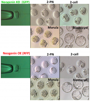
Phase-contrast images of embryos at different developmental stages via neogenin expression.[1]
--Mark Hill (talk) 16:17, 21 August 2014 (EST) This is all correct. The image is very large (4.8 MB), perhaps a small version could have been uploaded. You can adjust the resolution and size in most image editing programs.
Lab 3
Parathyroid gland
<pubmed>22808183</pubmed> <pubmed>22649358</pubmed> <pubmed>21881196</pubmed> <pubmed>21904825</pubmed>
Thymus
<pubmed>21733645</pubmed> <pubmed>20836742</pubmed> <pubmed>21263742</pubmed> <pubmed>512270</pubmed>
Pancreas
<pubmed>22761699</pubmed> <pubmed>22893718</pubmed> <pubmed>24496309</pubmed> <pubmed>24595965</pubmed> <pubmed>23822675</pubmed> <pubmed>22968764</pubmed>
Lab 4
1. Identify a paper that uses cord stem cells therapeutically and write a brief (2-3 paragraph) description of the paper's findings.
Regulation of Glioblastoma Progression by Cord Blood Stem Cells Is Mediated by Downregulation of Cyclin D1
Glioblastoma multiforme (GBM) is known to belong to a very life threatening form of brain cancer. Research is currently focused on finding treatments regarding such abnormalities, such as the application of neuronal stem cells in reducing the population of tumours, however there were many problems that occurred with such treatment. Recently, Human umbilical cord blood derived stem cells (hUCBSC) has been extensively used as they are useful mesenchymal stem cells that are easy to isolate and are more available. GBM is caused by the overexpression of cyclin D1 and its subsequent binding to Cdk 4/Cdk 6 which defines the rate limiting step required for the cell to progress further on to the cell cycle from the G1 phase. In order to stop this over expression, scientists have used hUCBSC to inhibit the cell from progressing on with the cell cycle.
When (hUCBSC) were cultured with U251 and 5310 cells, flow cytometry technology revealed that the cells underwent G1 arrest showing an increase in the G0-G1/S phase ratio. There was also a 54% reduction in levels of cyclin D1 when hUCBSC was cultured with U251 in comparison to the control. Immunoprecipiation revealed that hUCBSC treated cells when immuno blotted with Cdk 4 and Cdk 6 antibodies, down regulated expression of both Cdk 4 and Cdk 6. Western blot also showed the same down regulating pattern of the individual expression and genes which confirmed that there was cell cycle arrest, thus preventing tumours from forming.
As such, this study helps researchers to grasp the foundation of using hUCBSC as a treatment for glioblastoma and to further build on such research. Since it is evident that hUCBSC is effective in reducing cyclin D1 expression; analysing glioblastomal hierarchy will aid in providing the missing links needed to create the clinical treatment.
<pubmed>21455311</pubmed>
2. There are a number of developmental vascular "shunts" present in the embryo, that are closed postnatally. Identify these shunts and their anatomical location.
Three major vascular shunts include:
• Ductus venosus - is a shunt of oxygenated blood from umbilical vein to IVC, bypassing the liver. The ductus venosus constricts and closes soon after birth and becomes the ligamentum venosum
• Foramen ovale - is a flap valve in the atrial septum between the right and left atrium that shunts highly oxygenated blood . The remnant of the foramen ovale is known as the fossa ovalis.
• Ductus arteriosus - is a shunt from the descending aorta to the left pulmonary artery near the bifurcation of the pulmonary trunk. Permanent closure takes 4-6 weeks by fibrosis, and the remnant is referred to as the ligamentum arteriosum.
Lab 5
Select an abnormality of either gastrointestinal or respiratory development and write a brief description of developmental causes(s) for this abnormality. Your answer should be added to your own student page, be brief (2-3 paragraphs) and referenced.
Midgut volvulus
The embryonic development of the midgut has a number of steps which ensures its proper formation. These include the viability of the superior mesenteric artery to divide the midgut into the cephalad (pre-arterial region) and caudad (post-arterial region). [1]During the fourth gestational week, the gastrointestinal system is composed of a centrally positioned linear tube in the abdomen. At approximately 6 weeks of gestational age, the midgut undergoes a process of U-shaped herniation causing the two portions of the midguts to face the opposite directions in relation to the superior mesenteric artery. From this moment, a number of rotation events occur to ensure the complete development of the gastrointestinal tract as it becomes set in the posterior abdominal wall.[2]
In some cases malrotations can occur during the developmental process of the midgut which involves the complete twisting of the midgut in relation to the axis of the superior mesenteric artery. In extreme cases this can lead to midgut volvulus which results in a narrowed mesenteric base that can obstruct the passage of blood and lead to tissue necrosis. Other complications resulting from midgut volvulus include intestinal ischaemia, peritonitis, mucosal necrosis and sepsis which can eventually lead to death if left untreated. [3]
Malrotation is known to occur within 1 in 500 live births and out of those who develop midgut volvusos, 68-71% are neonates. [4]Although the actual cause of malrotation is unknown, researchers have made links to congenital syndromes such as Down syndrome and the VACTERL. It is also hypothesized that any embryonic interference during the normal patterns of rotation and fetal development can lead to midgut volvulus. Treatment of midgut volvulus is dependent on when the disease is diagnosed. [5]Normally a sigmoidoscopy is carried out as well as the Ladd procedure to resect dead gastrointestinal tissues. Transduodenal bands of ladd may also be divided to widen the mesenteric pedicle and prevent obstruction of blood flow. [6]Overall, research is still being conducted on the specific causes of malrotation and other numerous treatments.
Lab 6
Group work
Lab 7
1.Identify and write a brief description of the findings of a recent research paper on development of one of the endocrine organs covered in today's practical.
Novel genes upregulated when NOTCH signaling is disrupted during hypothalamic development.
It is known that the hypothalamus first develops from the ventral region of the diencephalon and signaling mechanisms such as the sonic hedgehog and bone morphogenic protein pathways are responsible for pattern arrangement. Neurogenesis is the key process required to ensure proper hypothalamus development which relies on many signaling pathways to produce neurons and glia. The Notch signaling pathway is currently known to inhibit neuronal differentiation and preserve neural progenitor identity. As a result of various studies and research, the combined theory has been implemented in this study to determine the effect of downregulating Notch signaling pathways in its effect on hypothalamic development.
Results show that when Notch signaling is inactivated, novel genes such as Dll1, Hes5, Hey1 and Ascl1 are upregulated in the rostral hypothalamus, subsequently leading to the early formation of hypothalamic neurons. This is also seen when embryos that are treated with DAPT (a chemical which inhibits cell differentiation mechanisms regulated by the Notch pathway) had an overexpression of cells differentiated into neurons in a clustered formation. This was compared to the control embryos which had differentiated cells in a scattered arrangement. As such, this research showed that Notch is a powerful signaling mechanism that is used to inhibit cell differentiation in order to control the number of cells differentiated into neurons or glia. This modulating ability of the Notch pathway is imperative in the early developing hypothalamus as it controls expression of cells and hence prevents any form of defects that can be harmful both prenatally and postnatally. Further research needs to be conducted on the Notch pathway to provide procedures where its mechanism can be used to resolve defects in the embryo and hence ensure proper hypothalamic development.
<pubmed>24360028</pubmed>
2.Identify the embryonic layers and tissues that contribute to the developing teeth.
Odontoblasts - neural crest-derived mesenchymal cells which establish the outer dental pulp. It differentiates via the enamel epithelium and releases dentin from dentinogenesis.
Ameloblasts - are derived from oral epithelium tissue of ectodermal origin and make up the inner enamel. They make pre-ameloblasts and produce enamel.
Periodontal ligament – is comprised of connective tissues which holds the tooth in place in the alveolar bone. It also encloses the cementum coating of the tooth root.
