User:Z3418837: Difference between revisions
No edit summary |
m (→Lab 2) |
||
| Line 70: | Line 70: | ||
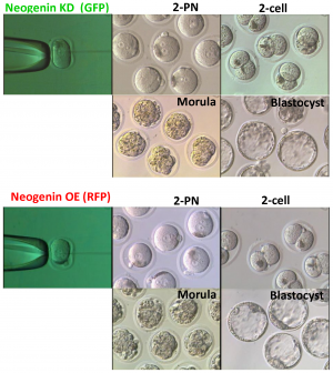
Phase-contrast images of embryos at different developmental stages via neogenin expression.<ref><pubmed>25013897</pubmed>| [http://www.plosone.org/article/info%3Adoi%2F10.1371%2Fjournal.pone.0101989 PLos One]</ref> | Phase-contrast images of embryos at different developmental stages via neogenin expression.<ref><pubmed>25013897</pubmed>| [http://www.plosone.org/article/info%3Adoi%2F10.1371%2Fjournal.pone.0101989 PLos One]</ref> | ||
--[[User:Z8600021|Mark Hill]] ([[User talk:Z8600021|talk]]) 16:17, 21 August 2014 (EST) This is all correct. The image is very large (4.8 MB), perhaps a small version could have been uploaded. You can adjust the resolution and size in most image editing programs. | |||
<references/> | <references/> | ||
Revision as of 16:17, 21 August 2014
Lab Attendance
- Lab 1 --Z3418837 (talk) 12:45, 6 August 2014 (EST)
- Lab 2 --Z3418837 (talk) 11:11, 13 August 2014 (EST)
- Lab 3 --Z3418837 (talk) 11:12, 20 August 2014 (EST)
- Lab 4
- Lab 5
- Lab 6
- Lab 7
- Lab 8
- Lab 9
- Lab 10
- Lab 11
- Lab 12
Individual Assessment
Lab 1
--Z3418837 (talk) 12:45, 6 August 2014 (EST)
http://www.ncbi.nlm.nih.gov/pubmed
<pubmed>25084016</pubmed>
Your Lab assessment now requires you to find a 2 recent research references on fertilisation or in vitro fertilisation. Paste each reference on your page, as shown in the class. Write below each reference a brief summary of the research article methods and findings. The summary for each need not be more than 3-4 paragraphs in length. This will need to be completed before next weeks laboratory.
Reference:PMID25077107
<pubmed>25077107</pubmed>
This study was designed to investigate whether the levels of vitamin D is an imperative factor when it comes to the clinical success of implantation and pregnancy rates in infertile women via invitro fertilisation.
Method summary
A cohort of 173 women were evaluated and selected for this study based on their age, follicle-stimulating hormone levels and their consent to undergo invitro fertilisation. The following study was conducted at Mount Sinai Hospital where proper facilities were available. Blood tests were then conducted for each patient to determine their levels of Vitamin D via the serum 25-hydroxy-vitamin D (25[OH]D) levels. Following the results of the blood test, the cohort were then categorised into two groups which was either vitamin D sufficient (≥ 75 nmol/L) or insufficient ( < 75 nmol/L) based on serum levels of 25(OH)D.
Each patient then underwent IVF cycles whereby standard agonists that contained the active ingredient 0.5 mg/d of buserelin acetate in conjunction with cetrolix acetate as the standard antagonist were used to control the length of the luteal phase and estradiol levels. The length and dose of the treatment were varied for each individual based on their demographic data. Serial transvaginal ultrasonograpy and serum lutenizing hormone assays were then used to check ovarian response. When 3 or more dominant follicles (≥ 17 mm) were produced, 10 000 IU of human chorionic gonadotropin was added to enhance nuclear maturation. Oocyte retrieval was then conducted via transvaginal ultrasound whereby it was fertilised and the resulting embryo was transferred 3-5 days post-fertilisation. The rate of pregnancy per IVF cycle was then used as the primary outcome for this study whereby the visibility of the intrauterine sac of the embryo determined implantation.
Results summary
Out of the 182 patients that participated in this study, it was found that only 173 patients could continue on with this trial as they satisfied the criteria, however only 162 were fit for embryo transfer. Following the results from the blood test, it was noted that 53.8% of patients had insufficient levels of 25(OH)D and 45.1% had sufficient amounts. It was discovered that 71.8% of those with sufficient levels of 25(OH)D were more likely to proceed with embryo transfer on day 5 compared to 58.9% (p = 0.054) of those assigned to the ‘insufficient 25(OH)D’ category. Other factors such as oocyte retrieval and frequency of intracytoplasmic sperm injection were fairly similar in both groups. The study revealed that there was a higher clinical pregnancy rate per IVF cycle for those assigned to the sufficient 25(OH)D level category by 52.5% compared to those with insufficient amounts of 25(OH)D which was 34.7% (p < 0.001). Similarly, there was a significant clinical pregnancy rate per embryo transfer of 54.7% in comparison to 37.9% in woman belonging to the sufficient and insufficient category respectively. It was also noted that the implantation rate was greater in the sufficient category compared to the insufficient group, however the difference was only minimal (p= 0.6). Overall, the results suggest that serum 25(OH)D levels may be a predictor of clinical pregnancy.
Reference:PMID24672163
<pubmed>24672163</pubmed>
The process of achieving pregnancy via invitro fertilisation needs to be monitored and controlled with respect to the demographics of the individual in order to achieve a successful outcome. This study focuses on predicting the value of β-human chorionic gonadotrophin (β-HCG) that can lead to clinical success.
Method summary
Data analysis was taken from 171 female patients using the statistical package for social sciences program whereby all IVF cycles were monitored. The cycles that showed fresh multi-cell embryos (day 3) or blastocysts (day 5) were deemed fit for the trial and were the ones that were further used in this study. Serum β-HCG concentrations were then taken 14 days after the embryo was transferred whereby a second test was done on day 16 only if the first test revealed a positive β-HCG result. This was done to predict values that could enable doctors to evaluate a healthy intrauterine pregnancy or a problematic ectopic pregnancy. After 6-7 weeks of pregnancy, ultrasounds were conducted to check cardiac activity as well as the amount of gestational sacs. This was then repeated at 12 weeks of pregnancy to ensure there was no chance of abortion.
A continuing pregnancy was defined as one that continued for at least 12 weeks of gestation and showed signs of proper cardiac function. On the other hand, those pregnancies that were abnormal had dropped levels of β-HCG concentrations and led to empty gestational sacs that showed no embryonic cardiac function. As a measuring tool for detecting levels of β-HCG, Chemiluminescent microparticle immunoassays were used whereby the measuring range established was between 0.0-15,000 mIU/mL. HCG levels above 10 IU/L signified early pregnancy.
Results Summary
Out of the 171 patients that participated in this study, only 139 were included due to the missing data on the levels of β-HCG concentrations at day 14 and 16 post embryonic transfer. In total there were 39 abnormal pregnancies that involved ectopic pregnancy, abortions and biochemical pregnancies (sufficient HCG levels detected but no visible gestational sac). Overall the patients were categorised into two groups which were patients with ‘ongoing pregnancy’ (n=100) and ‘without ongoing pregnancy’ (n=39). The Mann-Whitney test (statistical testing) was then used to compare the levels of β-HCG levels in both groups. It was found that the group with ongoing pregnancy had a median serum β-HCG level of 600 mIU/ml, whereas the other group had a median serum β-HCG level of 178 mIU/ml. This indicated a significant difference of P < 0.05 when comparing the two groups. It was also found that when serum β-HCG levels reached 347 mIU/ml, there was a 73.6% chance that the pregnancy was ongoing. Furthermore, there was no definite correlation established between age and the rate of ongoing pregnancy as both categories had patients of similar age groups with a combined range of 23-41 year old patients. Overall, the study revealed that early serum β-HCG is a potential predictor of successful outcomes in invitro fertilisation.
Lab 2
Phase-contrast images of embryos at different developmental stages via neogenin expression.[1]
--Mark Hill (talk) 16:17, 21 August 2014 (EST) This is all correct. The image is very large (4.8 MB), perhaps a small version could have been uploaded. You can adjust the resolution and size in most image editing programs.
