User:Z3331556: Difference between revisions
(→Lab 7) |
|||
| Line 13: | Line 13: | ||
--[[User:Z3331556|z3331556]] 11:52, 15 September 2011 (EST) | --[[User:Z3331556|z3331556]] 11:52, 15 September 2011 (EST) | ||
==Lab 1== | ==Lab 1== | ||
Revision as of 13:03, 21 September 2011
Attendance
--Z3331556 12:55, 28 July 2011 (EST)
--z3331556 11:57, 4 August 2011 (EST)
--z3331556 12:14, 11 August 2011 (EST)
--z3331556 11:09, 18 August 2011 (EST)
--z3331556 12:30, 25 August 2011 (EST)
--z3331556 11:16, 1 September 2011 (EST)
--z3331556 11:52, 15 September 2011 (EST)
Lab 1
1. Identify the origin of In Vitro Fertilization and the 2010 nobel prize winner associated with this technique.
The first successful IVF occurred in the UK in 1978 and Robert G. Edwards was awarded the Nobel Prize for this technique in 2010.Lecture - 2011 Course Introduction
2. Identify a recent paper on fertilisation and describe its key findings.
"Improved pregnancy rate with administration of hCG after intrauterine insemination: a pilot study" by Ilkka Y Järvelä, Juha S Tapanainen and Hannu Martikainen. Published on 23 February 2010 by Reproductive Biology and Endocrinology journal. They found that Intrauterine insemination (IUI), a common fertility treatment, improved pregnancy rate when hCG (human chorionic gonadotrophin)was administered after instead of before IUI. Pregnancy rates were 10.9% when hCG was given before IUI and 19.6% when hCG was given after IUI.[1]
3. Identify 2 congenital anomalies.
-Trisomy 21 (Down Syndrome) occurs when an extra copy of chromosome 21 is present
-Myelodysplasia (Spina bifida) is a condition where the fetus' spin fails to close in the first few months of pregnancy
--Mark Hill 10:10, 3 August 2011 (EST) These answers are fine.
Lab 2
1. Identify the ZP protein that spermatozoa binds and how is this changed (altered) after fertilisation.
The ZP protein that spermatozoa binds to is ZP3, when this occurs an Acrosome Reaction results where the head of the spermatozoa releases enzymes from the acrosomal vesicle (via exocytosis) which digests this protective coating of the egg (ZP3)[2] and exposes ZP2 to surface proteins of sperm Lecture - Fertilization
2.Identify a review and a research article related to your group topic.
PRIMARY ARTICLE
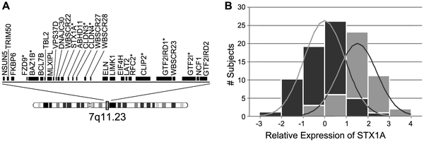
PLoS One. 2010 Apr 21;5(4):e10292. Intelligence in Williams Syndrome is related to STX1A, which encodes a component of the presynaptic SNARE complex. Gao MC, Bellugi U, Dai L, Mills DL, Sobel EM, Lange K, Korenberg JR. Source
Medical Genetics Institute, Cedars-Sinai Medical Center, Los Angeles, California, United States of America.
Abstract Although genetics is the most significant known determinant of human intelligence, specific gene contributions remain largely unknown. To accelerate understanding in this area, we have taken a new approach by studying the relationship between quantitative gene expression and intelligence in a cohort of 65 patients with Williams Syndrome (WS), a neurodevelopmental disorder caused by a 1.5 Mb deletion on chromosome 7q11.23. We find that variation in the transcript levels of the brain gene STX1A correlates significantly with intelligence in WS patients measured by principal component analysis (PCA) of standardized WAIS-R subtests, r = 0.40 (Pearson correlation, Bonferroni corrected p-value = 0.007), accounting for 15.6% of the cognitive variation. These results suggest that syntaxin 1A, a neuronal regulator of presynaptic vesicle release, may play a role in WS and be a component of the cellular pathway determining human intelligence.
PMID:20422020 [3]
- Williams Syndrome presents with a distinct pattern of intellectual disabilities that differ from normal on subtests of the WAIS-R (Wechsler Adult Intelligence Scale-Revised). Found that relative to their overall performance, WS subjects tended to do well in tests of vocabulary (Vocabulary) and abstract reasoning (Similarities, Picture Arrangement), and poorly in tests of numeracy (Arithmetic), visual-spatial (Digit Symbol, Block Design, Object Assembly), and memory (Digit Span)
- Gene expression in the tissue of interest (brain) is not possible so quantitated gene expression in lymphoblastoid (LB) cell lines
- STX1A is best known as an important component of the presynaptic SNARE complex involved in priming of synaptic vesicles for release.
- Data indicate that peripheral STX1A expression levels measured in lymphoblastoid cell lines strictly grown, is related to an emergent property of the CNS, intelligence.[4]
REVIEW ARTICLE
Arch Pediatr. 2009 Mar;16(3):273-82. Epub 2008 Dec 18. [Williams-Beuren syndrome: a multidisciplinary approach]. [Article in French] Lacroix A, Pezet M, Capel A, Bonnet D, Hennequin M, Jacob MP, Bricca G, Couet D, Faury G, Bernicot J, Gilbert-Dussardier B. Source
Laboratoire langage, mémoire et développement cognitif, CNRS, UMR 6215, 99, avenue du Recteur-Pineau, 86000 Poitiers, France. agnes.lacroix@uhb.fr
Abstract Williams-Beuren syndrome (WBS) (OMIM# 194050) is a rare, most often sporadic, genetic disease caused by a chromosomal microdeletion at locus 7q11.23 involving 28 genes. Among these, the elastin gene codes for the essential component of the arterial extracellular matrix. Developmental disorders usually associate an atypical face, cardiovascular malformations (most often supravalvular aortic stenosis and/or pulmonary artery stenosis) and a unique neuropsychological profile. This profile is defined by moderate mental retardation, relatively well-preserved language skills, visuospatial deficits and hypersociability. Other less known or rarer features, such as neonatal hypercalcemia, nutrition problems in infancy, ophthalmological anomalies, hypothyroidism, growth retardation, joint disturbances, dental anomalies and hypertension arising in adolescence or adulthood, should be treated. The aim of this paper is to summarize the major points of WBS regarding: (i) the different genes involved in the deletion and their function, especially the elastin gene and recent reports of rare forms of partial WBS or of an opposite syndrome stemming from a microduplication of the 7q11.23 locus, (ii) the clinical features in children and adults with a focus on cardiovascular injury, and (iii) the specific neuropsychological profile of people with WBS through its characteristics, the brain structures involved, and learning.
PMID:19097873 [5]
Lab 3
1. What is the maternal dietary requirement for late neural development?
Iodine is an important maternal dietary requirement for late neural development as a severe deficiency of this mineral during pregnancy seriously influences fetal brain development and in the worst case leads to cretinism, a decreased thyroid hormone production that has multiple complications. Recent studies have shown that even a mild iodine deficiency during pregnancy and during the first years of life adversely affects brain development. The World Health Organisation (WHO) considers iodine deficiency as the most common preventable cause of early childhood mental deficiency.[6] [7]
2. Upload a picture relating to you group project. Add to both the Group discussion and your online assessment page. Image must be renamed appropriately, citation on "Summary" window with link to original paper and copyright information. As outlined in the Practical class tutorial.
Lab 4
1. The allantois, identified in the placental cord, is continuous with what anatomical structure?
The allantois of the placental cord is an extra-embryonic membrane, that originates from the endodermal layer of the trilaminar embryo and extends from the early hindgut. Placenta Development
2. Identify the 3 vascular shunts, and their location, in the embryonic circulation
- Ductus venosus - between the umbilical vein and the inferior vena cava
- Foramen ovale - between the right and left atrium
- Ductus arteriosus - between the pulmonary artery and descending aorta
These shunts redirect oxygenated blood away from the lungs, liver and kidneys as these major organ's functions are run by the placenta at this point of the fetus' development Intermediate - Vascular Overview
3. Identify the Group project sub-section that you will be researching
Introduction
History of the disease
Etiology
Diagnosis
Genetic Factors
Physical Characteristics
Associated medical conditions
Cognitive, Behavioural and Neurological Problems
Epidemiology
Management/treatment
Specialized Facilities/ supportive associations
Case studies
Interesting facts
Current research and developments
Lab 5
1. Which side (L/R) is most common for diaphragmatic hernia and why?
Approximately 70 to 90% of Diaphragmatic hernias are 'Bochdalek-type,' or posterolateral hernias, most often occurring on the left posterolateral side. This is because the left pleuro-peritoneal canal is larger than the right, and therefore closing of this side occurs slightly later, hence more chance of hernia occurring on this side. [8] [9]
Lab 6
1. What week of development do the palatal shelves fuse?
The palatal shelves fuse in week 9 of development. This process requires the a growth and elevation of the palatal shelves before fusing in the midline Lecture - Head Development
2. What animal model helped elucidate the neural crest origin and migration of cells?
Chicken embryo model. Neural crest development has been best studied in avian embryos as they can be subject to "surgical manipulation, cell marking techniques, cell culture, and transgenesis by electroporation and retrovirally mediate gene transfer" [10]
3. What abnormality results from neural crest not migrating into the cardiac outflow tract?
Cranial neural crest cells extending from the auditory placode to somite 3 migrate to the outflow tract of the heart to participate in aorticopulmonary and truncal septation in the chick embryo. Surgical removal of these premigratory cells results in a high incidence of persistent truncus arteriosus [11] Failure of the outflow septum to form results in persistent truncus arteriosus, a condition in which there is a single outflow vessel with a single valve. [12]
Lab 7
1. Are satellite cells (a) necessary for muscle hypertrophy and (b) generally involved in hypertrophy?
Satellite cells are generally involved in muscle hypertrophy but they are not necessary
2. Why does chronic low frequency stimulation cause a fast to slow fibre type shift?
Chronic Low Frequency Stimulation (CLFS) is a standard, reproducible model of muscle training that parallels the stimulation of slow-twitch muscles by slow motoneurons. This artificial type of nerve innervation induces the sequential transitions in myosin heavy chain (MHC)expression, ultimately resulting in the transition of fast twitch to slow twitch fibres. [13]

