User:Z3294943
Attendance
--z3294943 11:06, 11 August 2011 (EST)-
-z3294943 11:12, 4 August 2011 (EST)
--z3294943 11:05, 18 August 2011 (EST)
--z3294943 11:05, 25 August 2011 (EST)
--z3294943 12:04, 1 September 2011 (EST)
--z3294943 11:08, 15 September 2011 (EST)
Online Assessment
LAB 1
- Identify the origin of In Vitro Fertilization and the 2010 nobel prize winner associated with this technique.
In 1978, Louise Brown, the first IVF baby was born. In Vitro Fertilisation (IVF) aids couples who may have trouble conceiving or are considered infertile. IVF involves the extraction of both the oocyte (female egg) and spermatozoa (male sperm) with fertilisation occurring outside the human body in a glass dish. Once fertilisation has occurred the zygote is then implanted back in to the female uterus. The idea of human IVF started in the mid 20th century by Robert G. Edwards who had studied fertilisation for many years and in 2010 Edwards was awarded the nobel prize in physiology or medicine. [1]
- Identify a recent paper on fertilisation and describe its key findings.
McAvey B Zapantis, A, Jindal SK, Lieman HJ, Polotsky AJ. (2011 ) How many eggs are needed to produce an assisted reproductive technology baby: is more always better? Fertility Sterility. 96(2):332-5. [2] This paper looked at the optimum number of oocytes that could be used in IVF to give the highest chance of live birth. They found 6-9 oocytes was advantageous in comparison to 5 or less oocytes, as well as, 10 or more oocytes. Thus suggesting more is not always better. This may aid future IVF patients not having to receive multiple treatments.
- Identify 2 congenital anomalies.
Congenital Anomalies are considered to be defects and/or disorders present at birth, such as Achondroplasia & Polydactyly.
--Mark Hill 00:43, 30 July 2011 (EST) Good the citation should be PMID:21718991 or I will show in lab how to generate a reference list.
LAB 2
- Identify the ZP protein that spermatozoa binds and how is this changed (altered) after fertilisation.
There are four Zona Pellucida (ZP) proteins within a human ZP1, ZP2, ZP3, ZP4. Before fertilization can occur the spermatozoa must undergo capacitation within the female uterus, in which the glycoprotein coat is detached from the spermatozoa's arcosomal surface. The zona pellucida surrounding the oocyte contains a glycoprotein ZP3, which acts as a receptor for the acrosome of the spermatozoa. Many spermatozoa release the contents of the acrosome, which lyse and degraded the zona pellucida thus exposing the surface of the oocyte to spermatozoa allowing it to bind to ZP2. The remaining zona pellucida undergoes a cortical reaction in which it increases the intracellular calcium levels causing depolarisation. This process allows the zona pellucida to become impermeable to other spermatozoa preventing polyspermy. ZP3 does undergo some changes after the cortical reaction it loses 1. its sperm binding ability 2. the capacity to promote the release of the spermatozoa's acrosome. [1] Occuring next would be the membrane fusion of the oocyte and spermatozoa.
- Identify a review and a research article related to your group topic. (Paste on both group discussion page with signature and on your own page)
- Review article PMID:11834588 This review looks at many different aspects of DMD and highlights how the lack of the gene dystrophin effects the brain, which may result in cognitive difficulties.
- Research articlePMID:20139167 This research article explores how branching of skeletal muscle causes weakness in the cyclic nature of regeneration and degredation in DMD.
References
- ↑ <pubmed>10341000</pubmed>
LAB 3
- What is the maternal dietary requirement for late neural development?
Iodine is a maternal dietary requirement for neural development with studies showing that it is a key source in providing thyroid associated hormones (T4) to the embryo before the thyroid of the foetus can take over. [1] If pregnant women become iodine deficient the foetus can suffer from hypothyroidism, goitres and abnormalities can arise, such as endemic cretinism. [2]
- Upload a picture relating to your group project
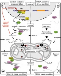
- Oxidative Stress Response in Friedreich Ataxia
--Mark Hill 15:30, 14 August 2011 (EST) This part is fine. I will be giving a tutorial in next week's lab to fix the referencing.
References
LAB 4
- The allantois, identified in the placental cord, is continuous with what anatomical structure?
The allantois in early embryonic life is an out-pouching of the hind gut and enters the connecting stalk. In later development the allantois becomes continuous with the developing superior urinary bladder and runs into the umbilicus. Though it is not a functional structure, the remnant can be seen in adults as the urachus, which extends from the apex of the bladder to the umbilicus creating the median umbilical ligament under the median periumbilical fold.
- Identify the 3 vascular shunts, and their location, in the embryonic circulation.
- Foramen Ovale: blood passes between the right to left atria
- Ductus Arteriosus: blood passes between the pulmonary trunk to the aorta
- Ductus Venosus: blood passes between umbilical vein to the inferior vena cava.
- Identify the Group project sub-section that you will be researching.
Introduction (history, epidemiology, age, gender, race) & Spinocerebellar physiology --z3294943 09:50, 24 August 2011 (EST)
LAB 5
- Which side (L/R) is most common for diaphragmatic hernia and why?
Congenital diaphragmatic hernia's (CDH) occur more commonly on the left side, approximately 90% of the time, more specifaclly at the foramen of Bochdalek. The posterolateral defect is due to incomplete fusion or abnormal formation of the pleuroperitoneal membranes. Reason as to why the left side is more commonly susceptible to CDH may be due to the fact that the right pleuroperitoneal membranes fuse much earlier then the left. --z3294943 17:00, 29 August 2011 (EST)
LAB 6
- What week of development do the palatal shelves fuse?
Palatogenesis occurs in two stages, primary and secondary palate development. During primary palatal development the embryonic palatal shelves are formed from mesenchyme of the medial nasal prominences and it continues to form the midline of the anterior maxilla. Secondary palatal fusion occurs in week 9 of a human embryonic development forming the hard and soft palates, which separate the nasal and oral cavities.
- What animal model helped elucidate the neural crest origin and migration of cells?
The chicken embryo provided visible migration of neural crest cells due to a Dil-label. The chick-quail chimera also provided great insight into the origin of neural crest cells. Nicole Le Douarin discovered that if a segment of neural tube and neural crest cells of a quail are implanted into a chick (at the the same embryonic stage) it is easy to observe the origin and migration of the neural crest cells.
- What abnormality results from neural crest not migrating into the cardiac outflow tract?
Neural crest cells contribute to the cardiac outflow tract, namely the aorta and pulmonary trunk, and are very important for cardiogenesis. If migration of these cells does not occur truncation and malformation of the aorticopulmonary septum can eventuate. This will lead to an aorticopulmonary septal defect in which there is communication between the aorta and pulmonary trunk via small aortic window. Furthermore, Tetralogy of Fallot is believed to arise from abnormal and/or absent cardiac neural crest migration. --z3294943 17:20, 11 September 2011 (EST)
LAB 7
- Are satellite cells (a) necessary for muscle hypertrophy and (b) generally involved in hypertrophy?
a) It has been shown that satellite cells are not necessary for muscle hypertrophy.
b) However, satellite cells will proliferate and differentiate during hypertrophy.
- Why does chronic low frequency stimulation cause a fast to slow fibre type shift?
Impulse activity is a determinant of fast and slow muscle fibre types. When chronic low frequency stimulation (CLFS) occurs on the nerves of fast muscle it can cause a phenotypical shift of the fast fibre type to a slow fibre type. [1] As slow type fibres generally make up the bulk of postural muscles, such as the erector spinae, they are receive low frequency stimulation to remain "switched on". So when fast muscle fibre type receive CLFS there a transition in the contractile properties of the muscle, namely myosin heavy chains, as well as the histochemical properties from a fast to a slow phenotype. [2]
References

