Talk:Basic - Primitive Heart Tube: Difference between revisions
No edit summary |
(→Content: Content added, not including animation) |
||
| Line 3: | Line 3: | ||
== Content == | == Content == | ||
--[[User:Z3212774|Phoebe Norville]] 12:23, 8 September 2009 (EST) Content for Section 1 of Basic Module: | |||
The heart is the first organ to function within an embryo. It starts to function at the beginning of the fourth week when the nutritional and oxygen requirements of the growing embryo can no longer be met by diffusion from the placenta. | |||
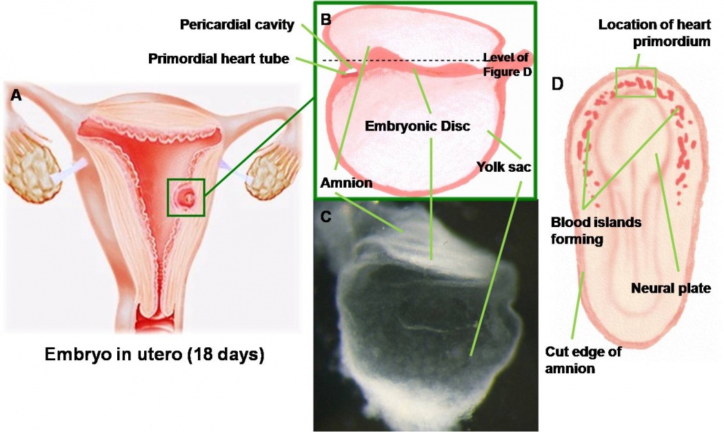
The heart initially forms from two tubes located bilaterally (on either side) of the trilaminar embryo in the cranial (head) region. The images below show these primitive tubes developing in an embryo approximately 18 days after conception. | |||
[[Image:HeartILP001.jpg|thumb|center|upright=3.5|The developing blood vessels and heart tube can be seen in an embryo at approximately 18 days]] | |||
[[Image:HeartILP002.jpg|thumb|upright=0.9|right|Fusion of the heart tubes]] | |||
===Embryonic Folding=== | |||
The disc-like embryo then undergoes a process of folding, in which both the cranial and lateral parts of the embryo fold ventrally (forwards). This brings the heart-forming region to a ventral (frontal) position. | |||
ANIMATION HERE. | |||
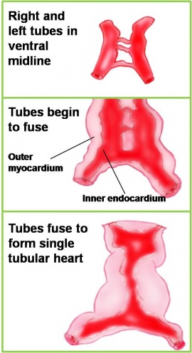
From here we can see the primitive heart from a ventral view as it consists of the two tubes. These tubes fuse together (as seen in the diagram on the right) to form a single, primordial heart tube, situated in the midline of the embryo, ventral to the pharynx. | |||
===Segments of the Heart Tube=== | |||
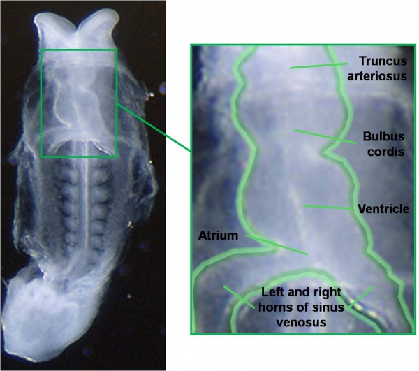
At this stage, the tube already has minor constrictions within it indicating sections of the heart tube that will form parts of the adult heart. The most caudal (tail end) segment of the heart tube is the sinus venosus which will later become the ends of the major veins carrying blood to the heart as well as parts of the atria. The next segments are the primitive atrium and primitive ventricle which will become the atria and ventricles of the adult heart. Cranial to these segments are the bulbus cordis, most of which will become the right ventricle, and the truncus arteriosus which forms the pulmonary and aortic trunks carrying blood away from the heart. | |||
[[Image:HeartILP-draft003.jpg|thumb|upright=2|center|22 day embryo showing segments of heart tube]] | |||
===Heart Tube Looping=== | |||
This tubular heart undergoes a process of looping during week four of development to form a shape that resembles that of the adult heart. | |||
[[Image:HeartILP-draft004.jpg|thumb|upright=4|center|Looping of the heart tube]] | |||
Revision as of 12:23, 8 September 2009
--Mark Hill 10:21, 7 September 2009 (EST) Outline seems fine. do you have in mind what these pictures will be?
Content
--Phoebe Norville 12:23, 8 September 2009 (EST) Content for Section 1 of Basic Module:
The heart is the first organ to function within an embryo. It starts to function at the beginning of the fourth week when the nutritional and oxygen requirements of the growing embryo can no longer be met by diffusion from the placenta.
The heart initially forms from two tubes located bilaterally (on either side) of the trilaminar embryo in the cranial (head) region. The images below show these primitive tubes developing in an embryo approximately 18 days after conception.
Embryonic Folding
The disc-like embryo then undergoes a process of folding, in which both the cranial and lateral parts of the embryo fold ventrally (forwards). This brings the heart-forming region to a ventral (frontal) position.
ANIMATION HERE.
From here we can see the primitive heart from a ventral view as it consists of the two tubes. These tubes fuse together (as seen in the diagram on the right) to form a single, primordial heart tube, situated in the midline of the embryo, ventral to the pharynx.
Segments of the Heart Tube
At this stage, the tube already has minor constrictions within it indicating sections of the heart tube that will form parts of the adult heart. The most caudal (tail end) segment of the heart tube is the sinus venosus which will later become the ends of the major veins carrying blood to the heart as well as parts of the atria. The next segments are the primitive atrium and primitive ventricle which will become the atria and ventricles of the adult heart. Cranial to these segments are the bulbus cordis, most of which will become the right ventricle, and the truncus arteriosus which forms the pulmonary and aortic trunks carrying blood away from the heart.
Heart Tube Looping
This tubular heart undergoes a process of looping during week four of development to form a shape that resembles that of the adult heart.


