Search results
From Embryology
Page title matches
- [[File:Developing_joint.jpg|thumb|Developing distal phalangeal joint]] ...lt, the region where two skeletal bones meet and articulate is called a "{{joint}}", that are classified based upon their: anatomical structure, mobility an17 KB (2,291 words) - 10:49, 14 February 2020
- '''Computational model of a synovial joint morphogenesis''' ...ggest that the mechanical and biochemical environments are crucial for the joint morphogenesis process.23 KB (3,350 words) - 23:48, 13 February 2020
Page text matches
- ...oint}} | {{musculoskeletal}} | {{cartilage}} | [[:Category:Joint|Category:Joint]] {| class="wikitable mw-collapsible mw-collapsed"564 bytes (59 words) - 23:12, 13 February 2020
- ...[[Musculoskeletal System - Skull Development|Skull Development]] | [[Head Development]]<noinclude>[[Category:Template]]</noinclude>747 bytes (83 words) - 11:31, 3 July 2015
- This {{Embryology}} category shows content related to knee development. ...endicular Skeleton]] | [[Book_-_Manual_of_Human_Embryology_11D|1910 - Limb Development]]7 members (0 subcategories, 0 files) - 14:07, 3 October 2017
- ...al joint]]<noinclude>[[Category:Template]][[Category:Term Link]][[Category:Joint]][[Category:Axial Skeleton]][[Category:Limb]][[Category:Cartilage]]</noincl226 bytes (25 words) - 23:11, 13 February 2020
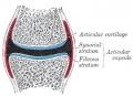
File:Gray0299.jpg ==Synovial Joint Cartoon== :'''Links:''' [[Musculoskeletal System - Joint Development|Joint Development]] | [[Cartilage Histology]](373 × 269 (34 KB)) - 07:44, 6 September 2011- ...nt|joint]]<noinclude>[[Category:Template]][[Category:Term Link]][[Category:Joint]][[Category:Axial Skeleton]][[Category:Limb]][[Category:Cartilage]]</noincl202 bytes (22 words) - 08:27, 17 April 2018
- ...etal]][[Category:Muscle]][[Category:Cartilage]][[Category:Bone]][[Category:Joint]]</noinclude>224 bytes (21 words) - 14:14, 6 May 2018
- ...ology}} | {{bone histology}} | [[Skeletal Muscle Histology]] | [[:Category:Musculoskeletal]] {{Historic Musculoskeletal links}}869 bytes (72 words) - 17:54, 27 July 2020
- ...{Embryology}} category shows pages and media related to the development of musculoskeletal joints. {{Joint Links}}58 members (1 subcategory, 32 files) - 11:29, 3 July 2015
- ...ry:Term Link]][[Category:Musculoskeletal]][[Category:Cartilage]][[Category:Joint]][[Category:Bone]]</noinclude>216 bytes (21 words) - 08:59, 20 November 2019
- ...ry:Term Link]][[Category:Musculoskeletal]][[Category:Cartilage]][[Category:Joint]][[Category:Bone]]</noinclude>211 bytes (21 words) - 09:56, 3 May 2018
- ...ted to both muscle (skeletal muscle), cartilage, bone (skeleton) and joint development. {{Musculoskeletal Links}}433 members (12 subcategories, 262 files) - 10:35, 11 April 2018
- ...is topic is closely linked to other topics: {{mesoderm}}, {{cartilage}}, {{joint}}, {{blood}}, {{skull}} {{Musculoskeletal Links}}442 members (4 subcategories, 276 files) - 10:18, 3 May 2018
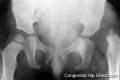
File:Congenital dislocation hip.jpg This X-ray shows incomplete development of the femur head within the pelvis joint. ...s|Limb Abnormalities]] | [[Musculoskeletal_System_-_Limb_Development|Limb Development]](400 × 265 (8 KB)) - 14:05, 22 May 2013- |+ colspan=2|[[Musculoskeletal System - Pelvis Development|'''Human Pelvis Development''']] | Sacroiliac joint forms.1 KB (154 words) - 00:47, 1 June 2018
- {{Ref-Ashley1955}} {| class="wikitable mw-collapsible mw-collapsed"2 KB (254 words) - 21:43, 13 November 2018
- {| class="wikitable mw-collapsible mw-collapsed" [[Talk:Mouse Development]] |6 KB (703 words) - 17:23, 27 March 2017
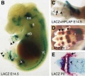
File:Joint development 02.jpg == Joint Development== (A) A 140-kb BAC from the Gdf5 locus was modified by inserting Cre-IRES-hPLAP into the translation start site of Gdf5 and used to make transgenic mi(454 × 403 (22 KB)) - 23:05, 21 March 2018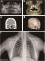
File:Cleidocranial dysplasia 01.jpg ...primary teeth, eruption failure of the permanent teeth, and impaired root development. ...lavicles and structural abnormalities occurring in the right shoulder peak joint.(518 × 700 (65 KB)) - 15:03, 13 February 2017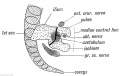
File:Keith1902 fig243.jpg ...in the ventral line. The cotyloid bone — os acetabuli — is formed in the Y-shaped cartilage between the three elements. It ossifies in the 13th year. P ...Musculoskeletal System - Pelvis Development|Pelvis Development]] | [[Fetal Development]](1,000 × 631 (98 KB)) - 12:26, 11 March 2018


