Uploads by Z5114433
From Embryology
This special page shows all uploaded files.
| Date | Name | Thumbnail | Size | Description | Versions |
|---|---|---|---|---|---|
| 15:57, 26 October 2017 | Purkinje Cell Arrangement.png (file) |  |
398 KB | =Purkinje Cell Arrangement= The modular organization of the cerebellum. Displays a series of "longitudinal sagittal bands" of Purkinje cell arrangement. Purkinje cell dendrites are flattened in the same direction as the microzones extend and are crosse... | 1 |
| 15:27, 26 October 2017 | Fetal MRI of cerebellum.png (file) |  |
260 KB | =Fetal MRI of cerebellum= Sagittal MRI of fetal cerebellum. Gradient echo of a fetus at 33 weeks of gestation. ==References== <pubmed>10.1177/0883073813486296</pubmed> | 1 |
| 14:44, 26 October 2017 | Flourens pigeon.gif (file) |  |
22 KB | =Flourens pigeon= A pigeon which had its brain lesioned in an one of Flourens initial experiments. This enabled Flourens to understand the gross anatomy of the cerebrum and cerebellum of mammals. ==References== http://www.cerebromente.org.br/n01/freno... | 1 |
| 14:33, 26 October 2017 | Ferrier's findings of cerebellum.gif (file) |  |
42 KB | =Ferrier's findings on a monkey's cerebellum with stimulation points= ==Reference== <ref>http://www.cerebromente.org.br/n01/frenolog/frenloc.htm</ref> ===Copyright=== | 1 |
| 12:48, 25 October 2017 | Second Trimester Cerebellum.jpeg (file) |  |
26 KB | =Second Trimester Cerebellum= Caudal, inferior image of the cerebellum at second trimester ==Reference== <ref>http://www.fetalultrasound.com/online/text/2-006.HTM</ref> ===Copyright=== | 1 |
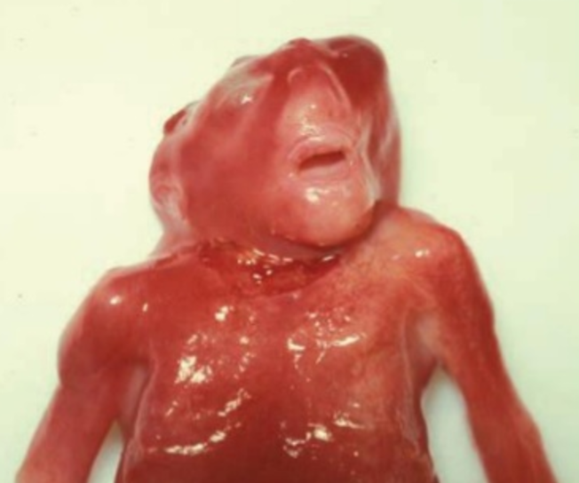
| 22:22, 4 October 2017 | Anencephaly.png (file) |  |
343 KB | =Anencephaly= An image of a stillborn baby due to an undeveloped brain and incomplete skull ==Reference== <ref>https://www.slideshare.net/NamXal1/development-of-nervous-system</ref> ===Copyright=== © 2014 ''Associated Press text, photo, graphic, au... | 1 |
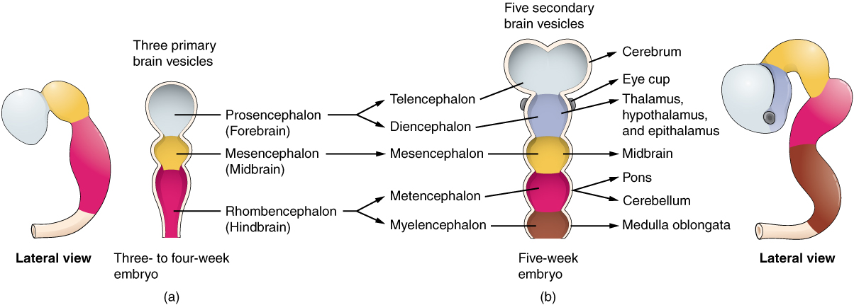
| 21:58, 4 October 2017 | Secondary brain vesicle.jpeg (file) |  |
209 KB | =Secondary brain vesicle = (Week 5) 3 primary vesicles develop into 5 secondary vesicles; - Prosencephalon develops into telencephalon (which includes the endbrain and cerebral hemispheres) and diencephalon (located between the brain and forms an opti... | 1 |
| 21:30, 4 October 2017 | Primary Brain Vesicles.jpeg (file) |  |
64 KB | =Primary Brain Vesicles= In week 4, 3 primary brain vesicles are formed; forebrain (prosencephalon) midbrain (mesencephalon), and hindbrain (rhombencephalon). These structures then differentiate into 5 secondary brain vesicles during week 5. ==Referen... | 1 |
| 16:52, 29 September 2017 | Arteries of the cerebellum.jpeg (file) |  |
51 KB | =Arteries of the cerebellum= | 1 |
| 16:10, 29 September 2017 | Cerebellum anatomical subdivisions.png (file) |  |
67 KB | =Superior view of anatomical subdivisions of the cerebellum= Superior view of the 3 cerebellar zones. The middle is the vermis. Either side of the vermis is the intermediate zone. Lateral to the intermediate zone is the lateral hemispheres. There is n... | 1 |
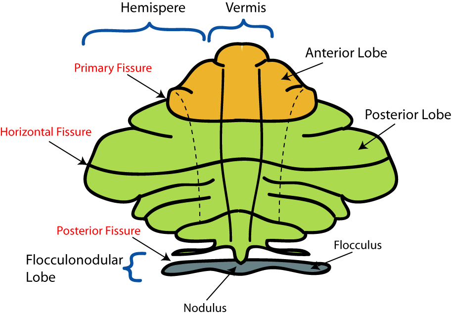
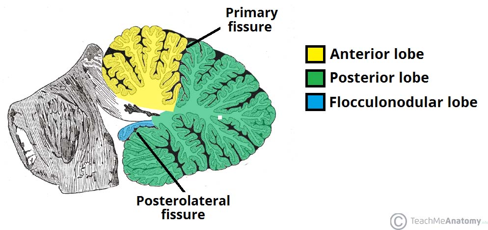
| 14:27, 29 September 2017 | Anatomical-Lobes-of-the-Cerebellum.jpg (file) |  |
102 KB | Anatomical lobe of the cerebellum The image depicts the anterior lobe which is rostral to the primary fissure, the posterior lobe (dorsal to the "primary fissure") and the flocculonodular lobe. | 1 |
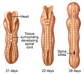
| 20:29, 14 September 2017 | Spina-bifida-lg-enlg - embryo.jpg (file) |  |
14 KB | Image of the Process of Spina Bifida during 21, 22 and 28 days. | 1 |
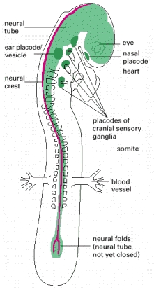
| 16:37, 14 September 2017 | Ch21f91.jpg (file) |  |
65 KB | Diagram of 2 day old embryo. The neural tube (green) is closed and the neural crest (red) is dorsal from ectoderm (above neural tube). | 1 |