Uploads by Z3333865
From Embryology
This special page shows all uploaded files.
| Date | Name | Thumbnail | Size | Description | Versions |
|---|---|---|---|---|---|
| 21:23, 4 October 2012 | Middle ear ossicles.png (file) |  |
914 KB | '''A view from inside the tympanic cavity of ''Lagenorhynchus obliquidens'' (Pacific White-sided Dolphin)''' This is a backlit view through the translucent floor of the ear trumpet (sulcus of mallear ridge). M = malleus; I = incus; sp = sigmoid proces | 1 |
| 21:05, 4 October 2012 | Semi-circular canals.jpg (file) |  |
98 KB | '''Wild-type semi-circular canal structure of zebrafish''' Three-dimensional reconstructions of adult wild-type zebrafish inner ears. C=lateral views of left hand ears, with anterior to the left. F=dorsal view. Scale bar, 500 µm. Abbrevations: a-amp, a | 1 |
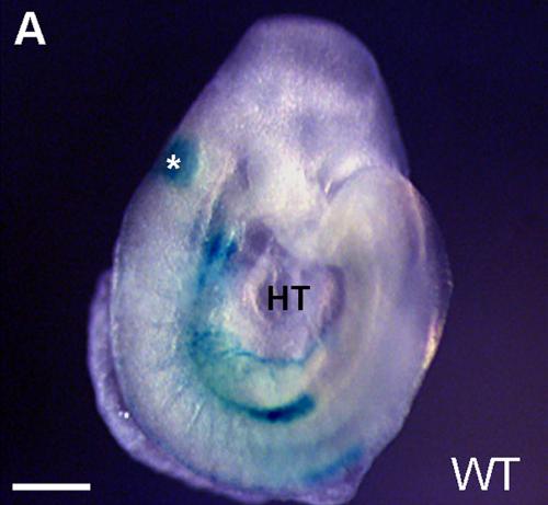
| 10:09, 3 October 2012 | Otic placode embryo.jpg (file) |  |
23 KB | '''Otic placode of an embryo''' The lateral view of an E8.5 embryo used as the control Hoxa3-lacZ (normal wild-type). Asterisk indicates the otic vesicle. HT, heart tube. Scale bars = 100 µm. Reference: <pubmed>22110697</pubmed> Copyright: © 2011 Di | 1 |
| 09:47, 3 October 2012 | Otic vesicle.jpg (file) |  |
24 KB | '''Zebrafish otic vesicle''' Otic vesicle morphology 27 hours post fertilisation. Reference: <pubmed>22164214</pubmed> Copyright: © 2011 Bhat, Riley. This is an open-access article distributed under the terms of the Creative Commons Attribution Licen | 1 |
| 09:33, 3 October 2012 | Cochlea stereocilia bundle.jpg (file) |  |
31 KB | '''Stereocilia bundle counts in the normal cochlea''' Examples of normal-looking stereocilia bundles. Modified from: Yang S-M, Chen W, Guo W-W, Jia S, Sun J-H, et al. (2012) '''Regeneration of Stereocilia of Hair Cells by Forced Atoh1 Expression in the | 1 |
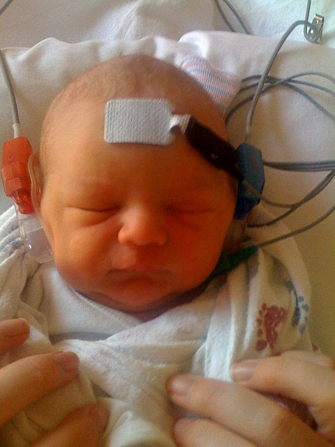
| 14:10, 2 October 2012 | Infant hearing test.jpg (file) |  |
82 KB | '''Testing the hearing of an infant''' Photo of Infant Hearing Test (Auditory Brainstem Response test) by Dom Sagolla, shared through flickr. Creative Commons copyright: Attribution-NonCommercial-NoDerivs CC BY-NC-ND This license is the most restric | 1 |
| 17:32, 29 September 2012 | Aquaporins.png (file) |  |
2.05 MB | '''Aquaporins''' Aquaporins 1, 4, and 5 in the inner ear (A-C) and middle ear (D-G). A: AQP1 in the inner ear showed positive staining of the lateral lining of the spiral ligament and some fibrocytes within the spiral ligament. B: AQP4 in the inner ear p | 1 |
| 16:53, 29 September 2012 | Atoh1 hair cell loss.png (file) |  |
4.47 MB | '''Conditional deletion of Atoh1 results in death of organ of Corti cells and patchy Myo7a-positive presumptive hair cells which are innervated by many nerve fibers.''' (a–b) Cell death assay using immunohistochemistry of activated Caspase 3 (red) and | 1 |
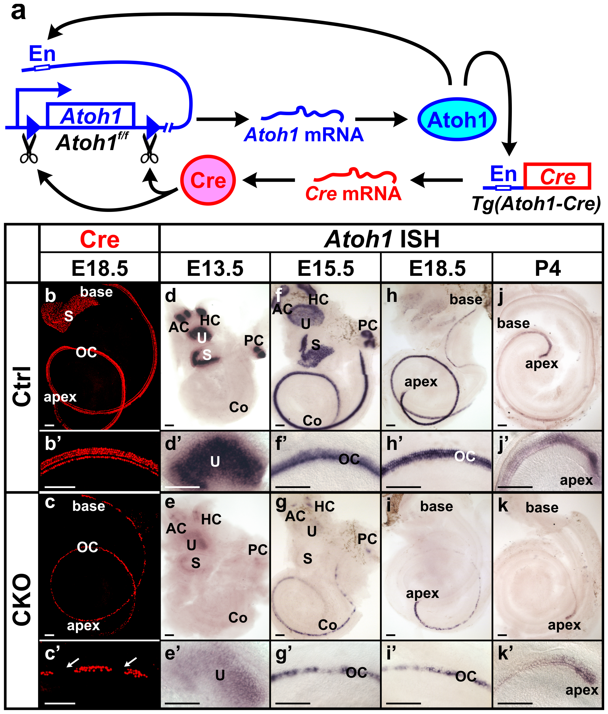
| 16:31, 29 September 2012 | Atoh1 model.png (file) |  |
3.5 MB | © 2012 Pan et al. This is an open-access article distributed under the terms of the Creative Commons Attribution License, which permits unrestricted use, distribution, and reproduction in any medium, provided the original author and source are credited. | 1 |
| 11:16, 19 September 2012 | Z3333865.gene expression.jpeg (file) |  |
51 KB | '''Representative expression patterns of genes controlling cochlear and vestibular specification.''' As described in the review by Chatterjee ''et al.'' (2010): (A) Shh functions to maintain Pax2 and restrict Dlx5/Dlx6 in the medial wall of the otic ve | 1 |
| 11:10, 19 September 2012 | Z3333865.gene expression.pdf (file) | 158 KB | '''Representative expression patterns of genes controlling cochlear and vestibular specification.''' As described in the review by Chatterjee ''et al.'' (2010): (A) Shh functions to maintain Pax2 and restrict Dlx5/Dlx6 in the medial wall of the otic ve | 1 | |
| 13:11, 14 September 2012 | Neural fate.jpg (file) |  |
29 KB | Student Image. Based upon the text provided by <pubmed> | 1 |
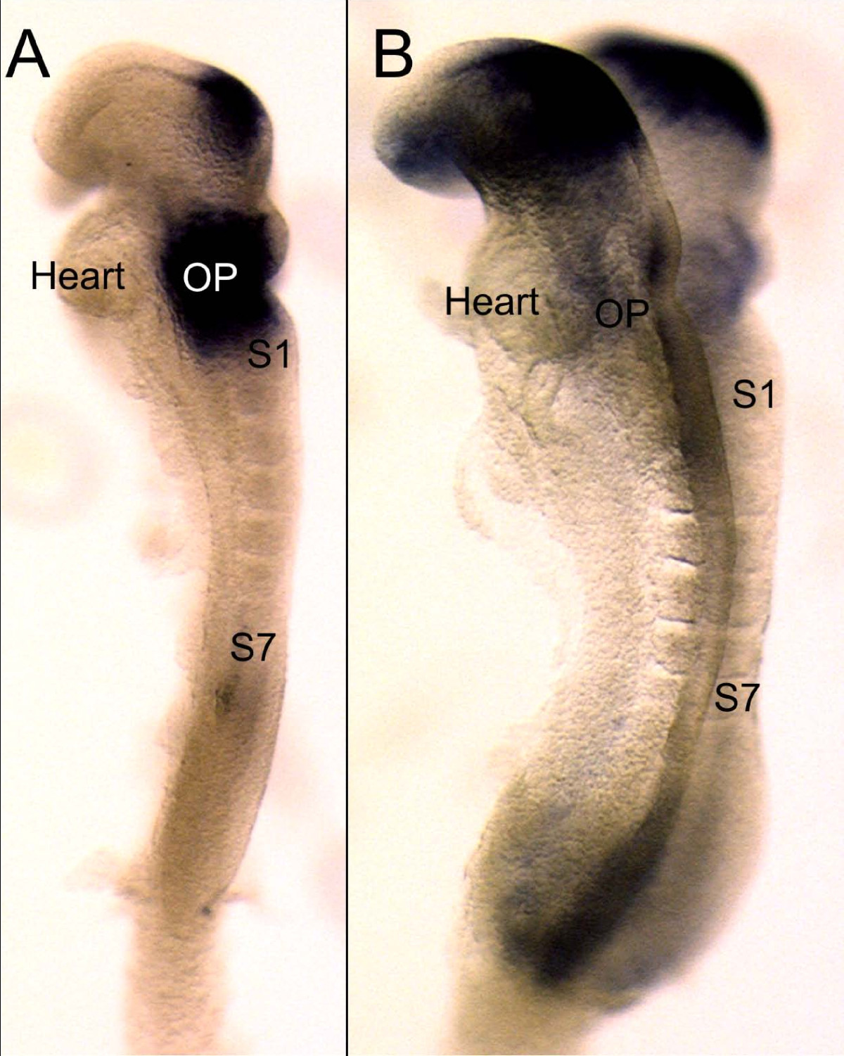
| 13:29, 11 September 2012 | Z3333865.pax2 and 8.jpg (file) |  |
339 KB | '''Early expression of Pax2 and Pax8 compared''' This image shows the earliest expression of Pax8 (A) and Pax2 (B) in 7 somite (S1, S7) mouse embryos of approximately 8 embryonic days. Note that at this stage Pax8 is more profoundly expressed throughout | 1 |
| 12:35, 11 September 2012 | Z3333865.stages development inner ear.jpg (file) |  |
182 KB | '''Developmental milestones in mouse inner ear formation''' Competence of surface ectoderm lateral to both sides of the hindbrain (HB) precedes any cell morphology changes. (A) Thickening of surface ectoderm (SE) to form the early placodes (EP) which is | 1 |
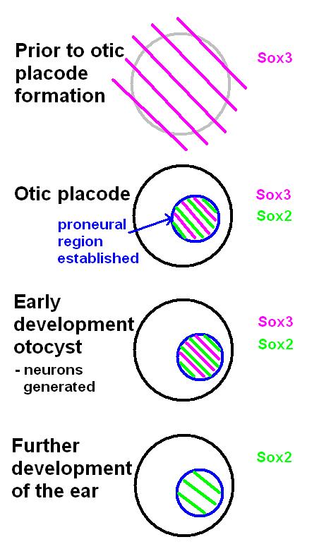
| 16:57, 7 September 2012 | Neural domain.jpg (file) |  |
54 KB | A schematic and simplistic representation of the expression of Sox2 and Sox3 during development of the ear. Before the otic placode becomes distinct, Sox3 is already expressed in a broad area around the location of the future otic placode. Later in it wi | 1 |
| 10:13, 22 August 2012 | Normal cochlea.png (file) |  |
141 KB | Wild-type inner ear showing normal morphology. Structures are labeled as follows: aa, anterior ampulla; asc, anterior semicircular canal; cd, cochlea duct; ed, endolymphatic duct; la, lateral ampulla; lsc, lateral semicircular canal; pa, posterior ampul | 1 |
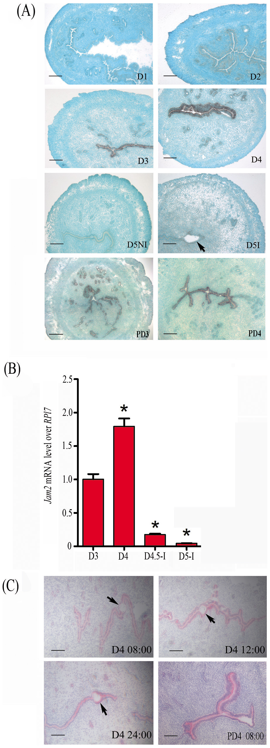
| 14:37, 4 August 2012 | Z3333865.implantation.png (file) |  |
2.87 MB | '''Jam2 expression in mouse uterus during early pregnancy.''' (A) In situ hybridization of Jam2 mRNA. (B) Real-time RT-PCR quantification of Jam2 mRNA. (C) JAM2 immunostaining. D1, day 1; D2, day 2; D3, day 3; D4, day 4; D4.5-I, implantation site at day | 1 |
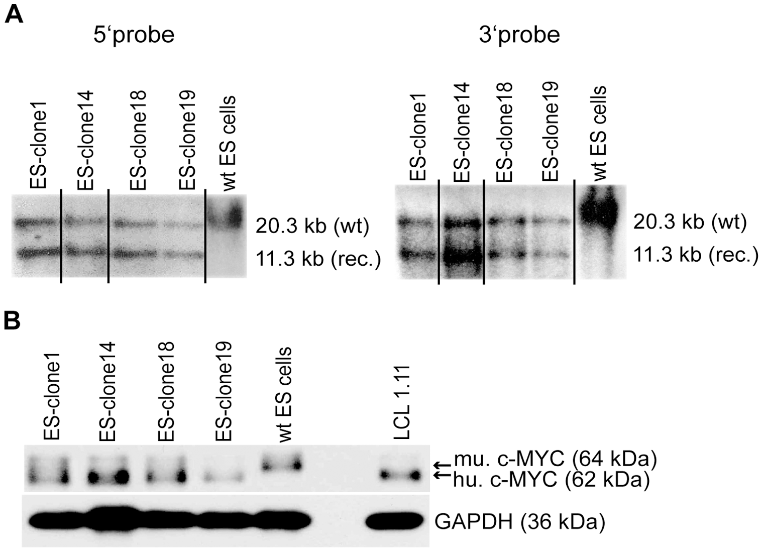
| 12:04, 1 August 2012 | Z3333865.homologous recombination and c-MYC2 expression in ES cell clones.png (file) |  |
282 KB | '''Confirmation of homologous recombination and c-MYC2 expression in ES cell clones.''' (A) Genomic DNA of ES cell clones 1, 14, 18 and 19 and of wildtype ES cells (wt Bruce 4) was digested with EcoRI. Digested DNA was analyzed by Southern blotting with | 1 |