Quicktime Movie - CT Mouse Face: Difference between revisions
No edit summary |
m (Redirected page to Mouse Stages Face microCT Movie) |
||
| (7 intermediate revisions by the same user not shown) | |||
| Line 1: | Line 1: | ||
#REDIRECT [[Mouse_Stages_Face_microCT_Movie]] | |||
{| border='0px' | {| border='0px' | ||
|- | |- | ||
| <qt>file=Mouse_face_microCT_01.mov|width=240px|height= | | <qt>file=Mouse_face_microCT_01.mov|width=240px|height=380px|controller=true|autoplay=false</qt> | ||
| valign="top" |[[File:Mouse_face_microCT_01.jpg| | |||
Unlabeled version | |||
| valign="top" |[[File:Mouse_face_microCT_01.jpg|160px]] | |||
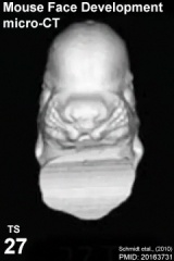
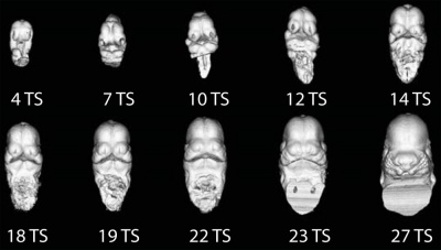
Ventral view of the developing mouse face (E9.5 - E12) the series of μCT scans showing the range of shape and size variation. | Ventral view of the developing mouse face (E9.5 - E12) the series of μCT scans showing the range of shape and size variation. | ||
| Line 9: | Line 12: | ||
'''Links:''' [[Media:Mouse_face_microCT_01.mov|Quicktime]] | [[Quicktime Movie - CT Mouse Face|Quicktime version]] | [[Movie - CT Mouse Face|Flash version]] | [[Mouse Development]] | '''Links:''' [[Media:Mouse_face_microCT_01.mov|Quicktime]] | [[Quicktime Movie - CT Mouse Face|Quicktime version]] | [[Movie - CT Mouse Face|Flash version]] | [[Mouse Development]] | ||
See also movie of human face development between week 6 to 7 (Carnegie stage 16 to stage 18) [[Quicktime_Movie_-_Stage_16_to_18_Face|Quicktime version]] | [[Movie_-_Stage_16_to_18_Face|Flash version]]. | |||
|- | |- | ||
| <qt>file=Mouse_face_microCT_02.mov|width=240px|height=380px|controller=true|autoplay=false</qt> | |||
Labeled version | |||
| [[File:Mouse_CT_E9.5-E12_head.jpg|400px]] | |||
* '''S''' - stomodeum | |||
* '''fnp''' - frontonasal prominence | |||
|} | |} | ||
| Line 26: | Line 39: | ||
--[[User:Z8600021|Mark Hill]] 13:27, 6 April 2012 (EST) Animation was constructed from individual images in panel A of [http://www.biomedcentral.com/1471-213X/10/18/figure/F1 figure 1.] | --[[User:Z8600021|Mark Hill]] 13:27, 6 April 2012 (EST) Animation was constructed from individual images in panel A of [http://www.biomedcentral.com/1471-213X/10/18/figure/F1 figure 1.] | ||
Latest revision as of 12:42, 10 May 2013
Redirect to:
| width=240px|height=380px|controller=true|autoplay=false</qt>
Unlabeled version |

Ventral view of the developing mouse face (E9.5 - E12) the series of μCT scans showing the range of shape and size variation.
Links: Quicktime | Quicktime version | Flash version | Mouse Development
|
| width=240px|height=380px|controller=true|autoplay=false</qt>
Labeled version |

|
Reference
<pubmed>20163731</pubmed>| PMC2836989 | BMC Dev Biol.
BMC Dev Biol. 2010; 10: 18.
Published online 2010 February 17. doi: 10.1186/1471-213X-10-18
© 2010 Schmidt et al; licensee BioMed Central Ltd. This is an Open Access article distributed under the terms of the Creative Commons Attribution License (http://creativecommons.org/licenses/by/2.0), which permits unrestricted use, distribution, and reproduction in any medium, provided the original work is properly cited.
--Mark Hill 13:27, 6 April 2012 (EST) Animation was constructed from individual images in panel A of figure 1.