Notochord Movie: Difference between revisions
No edit summary |
No edit summary |
||
| Line 15: | Line 15: | ||
'''Links:''' [[Media:Notochord 02.mov|Quicktime version]] | [[Notochord]] | '''Links:''' [[Media:Notochord 02.mp4|MP4 version]] | [[Media:Notochord 02.mov|Quicktime version]] | [[Notochord]] | ||
|- | |- | ||
| width=250px|<mediaplayer width='240' height='200' image="http://embryology.med.unsw.edu.au/embryology/images/1/1e/Notochord_01_icon.jpg">Notochord 01.mp4</mediaplayer> | | width=250px|<mediaplayer width='240' height='200' image="http://embryology.med.unsw.edu.au/embryology/images/1/1e/Notochord_01_icon.jpg">Notochord 01.mp4</mediaplayer> | ||
| Line 31: | Line 31: | ||
'''Links:''' [[Media:Notochord 01.mov|Quicktime version]] | '''Links:''' [[Media:Notochord 01.mp4|PM4 version]] | [[Media:Notochord 01.mov|Quicktime version]] | [[Notochord]] | ||
|- | |- | ||
|} | |} | ||
Revision as of 16:03, 2 March 2013
| Embryology - 4 May 2024 |
|---|
| Google Translate - select your language from the list shown below (this will open a new external page) |
|
العربية | català | 中文 | 中國傳統的 | français | Deutsche | עִברִית | हिंदी | bahasa Indonesia | italiano | 日本語 | 한국어 | မြန်မာ | Pilipino | Polskie | português | ਪੰਜਾਬੀ ਦੇ | Română | русский | Español | Swahili | Svensk | ไทย | Türkçe | اردو | ייִדיש | Tiếng Việt These external translations are automated and may not be accurate. (More? About Translations) |
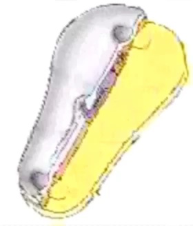
| <mediaplayer width='214' height='220' image="http://embryology.med.unsw.edu.au/embryology/images/9/91/Notochord_02_icon.jpg">Notochord 02.mp4</mediaplayer> | This is a dorsal view of the embryonic disc, caudal (tail and connecting stalk end) to the bottom and rostral (head end) to the top. The indentations show the location of the cloacal (bottom) and buccopharyngeal (top) membranes. The raised region in the middle of the embryonic disc is the primitive node (Hensen's node). The right hand side of the gastrulating embryonic disc is removed to the midline to show the the position of the initial axial process (purple). As the animation plays the axial process extends rostrally from the primitive node towards the buccopharyngeal membrane, where it stops. A cross-section view above the primitive node is shown in the second animation below.
|
| <mediaplayer width='240' height='200' image="http://embryology.med.unsw.edu.au/embryology/images/1/1e/Notochord_01_icon.jpg">Notochord 01.mp4</mediaplayer> |
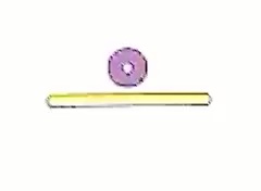
The view is a cross-section showing how the axial process initially is formed, then fused with the endoderm, to finally separate as a midline mesoderm structure.
Yellow - endoderm | Purple - axial process
Links: PM4 version | Quicktime version | Notochord |
Source: UNSW Embryology Thanks to the late Prof William Larsen for allowing permission to use animations based on images from his textbook. (More? movie technical information)
Links: Movies Page | Category:Movies
Cite this page: Hill, M.A. (2024, May 4) Embryology Notochord Movie. Retrieved from https://embryology.med.unsw.edu.au/embryology/index.php/Notochord_Movie
- © Dr Mark Hill 2024, UNSW Embryology ISBN: 978 0 7334 2609 4 - UNSW CRICOS Provider Code No. 00098G