Nodal Cilia Movie: Difference between revisions
mNo edit summary |
mNo edit summary |
||
| (2 intermediate revisions by the same user not shown) | |||
| Line 2: | Line 2: | ||
{| border='0px' | {| border='0px' | ||
|- | |- | ||
| width=360px|< | | width=360px|<html5media height="280" width="340">File:Nodal_cilia_001.mp4</html5media> | ||
[[Media:Nodal_cilia_001.mp4|'''Click Here''' to play on mobile device]] | |||
| valign="top" | | | valign="top" | | ||
===Cilia rotation an the mouse embryo (E 8.0) primitive node=== | ===Cilia rotation an the mouse embryo (E 8.0) primitive node=== | ||
| Line 35: | Line 36: | ||
Cilium cartoon | Cilium cartoon | ||
===Reference=== | ===Reference=== | ||
{{#pmid:16035921}} | |||
====Copyright==== | ====Copyright==== | ||
| Line 52: | Line 50: | ||
{{Footer}} | |||
[[Category:Mouse]] [[Category:Week 3]] | [[Category:Mouse]] [[Category:Week 3]] [[Category:Mouse E8.0]] | ||
Latest revision as of 09:23, 31 July 2018
| Embryology - 30 Apr 2024 |
|---|
| Google Translate - select your language from the list shown below (this will open a new external page) |
|
العربية | català | 中文 | 中國傳統的 | français | Deutsche | עִברִית | हिंदी | bahasa Indonesia | italiano | 日本語 | 한국어 | မြန်မာ | Pilipino | Polskie | português | ਪੰਜਾਬੀ ਦੇ | Română | русский | Español | Swahili | Svensk | ไทย | Türkçe | اردو | ייִדיש | Tiếng Việt These external translations are automated and may not be accurate. (More? About Translations) |
| <html5media height="280" width="340">File:Nodal_cilia_001.mp4</html5media> |
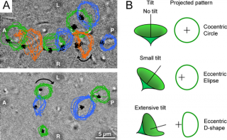
Cilia rotation an the mouse embryo (E 8.0) primitive nodeThe direction of cilia rotation was clockwise.
|

|
Note that in the mouse this occurs during week 1, in humans this occurs week 2 to 3 around gastrulation. |
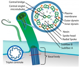
Cilium cartoon
Reference
Nonaka S, Yoshiba S, Watanabe D, Ikeuchi S, Goto T, Marshall WF & Hamada H. (2005). De novo formation of left-right asymmetry by posterior tilt of nodal cilia. PLoS Biol. , 3, e268. PMID: 16035921 DOI.
Copyright
© 2005 Nonaka et al. This is an open-access article distributed under the terms of the Creative Commons Attribution License, which permits unrestricted use, distribution, and reproduction in any medium, provided the original work is properly cited.
External Links Notice - The dynamic nature of the internet may mean that some of these listed links may no longer function. If the link no longer works search the web with the link text or name. Links to any external commercial sites are provided for information purposes only and should never be considered an endorsement. UNSW Embryology is provided as an educational resource with no clinical information or commercial affiliation.
Cite this page: Hill, M.A. (2024, April 30) Embryology Nodal Cilia Movie. Retrieved from https://embryology.med.unsw.edu.au/embryology/index.php/Nodal_Cilia_Movie
- © Dr Mark Hill 2024, UNSW Embryology ISBN: 978 0 7334 2609 4 - UNSW CRICOS Provider Code No. 00098G
