Nodal Cilia Movie: Difference between revisions
m (→Reference) |
mNo edit summary |
||
| Line 2: | Line 2: | ||
{| border='0px' | {| border='0px' | ||
|- | |- | ||
| width=360px|<mediaplayer width='340' height=260' image="http:// | | width=360px|<mediaplayer width='340' height=260' image="http://php.med.unsw.edu.au/embryology/images/a/a0/Node_cilia_movement.png">File:Nodal_cilia_001.mp4</mediaplayer> | ||
| valign="top" | | | valign="top" | | ||
| Line 20: | Line 20: | ||
|- | |- | ||
| [[File:Node cilia movement.png|320px]] | | [[File:Node cilia movement.png|320px]] | ||
| | | | ||
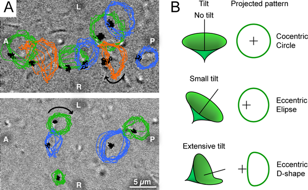
* '''A''' - Trace of node cilia in enhanced DIC images after background subtraction. Positions of root are indicated in black, and tip in blue, green, and orange. Most cilia have a pattern consistent with the projection of a tilted cone (blue and green, see text) whereas some cilia move in a D-shape (orange). A, P, L, and R refer to anterior, posterior, left, and right sides of the node, respectively. The direction of cilia rotation was clockwise (arrows). | |||
* '''B''' - Relationship between essentially rotatory movement of cilia and their projected images at various tilt angles. | |||
Note that in the mouse this occurs during week 1, in humans this occurs week 2 to 3 around [[Gastrulation|gastrulation]]. | |||
Note that in the mouse this occurs during week 1, in humans this occurs week 2 to 3 around gastrulation. | |||
|- | |- | ||
| Line 38: | Line 38: | ||
<pubmed>16035921</pubmed>| [http://www.plosbiology.org/article/info%3Adoi%2F10.1371%2Fjournal.pbio.0030268 PLoS] | <pubmed>16035921</pubmed>| [http://www.plosbiology.org/article/info%3Adoi%2F10.1371%2Fjournal.pbio.0030268 PLoS] | ||
====Copyright==== | ====Copyright==== | ||
Revision as of 22:37, 14 December 2013
| Embryology - 30 Apr 2024 |
|---|
| Google Translate - select your language from the list shown below (this will open a new external page) |
|
العربية | català | 中文 | 中國傳統的 | français | Deutsche | עִברִית | हिंदी | bahasa Indonesia | italiano | 日本語 | 한국어 | မြန်မာ | Pilipino | Polskie | português | ਪੰਜਾਬੀ ਦੇ | Română | русский | Español | Swahili | Svensk | ไทย | Türkçe | اردو | ייִדיש | Tiếng Việt These external translations are automated and may not be accurate. (More? About Translations) |
| <mediaplayer width='340' height=260' image="http://php.med.unsw.edu.au/embryology/images/a/a0/Node_cilia_movement.png">File:Nodal_cilia_001.mp4</mediaplayer> |
Cilia rotation an the mouse embryo (E 8.0) primitive nodeThe direction of cilia rotation was clockwise. A) Trace of node cilia in enhanced DIC images after background subtraction. Positions of root are indicated in black, and tip in blue, green, and orange. Most cilia have a pattern consistent with the projection of a tilted cone (blue and green, see text) whereas some cilia move in a D-shape (orange). A, P, L, and R refer to anterior, posterior, left, and right sides of the node, respectively. The direction of cilia rotation was clockwise (arrows). (B) Relationship between essentially rotatory movement of cilia and their projected images at various tilt angles.
|

|
Note that in the mouse this occurs during week 1, in humans this occurs week 2 to 3 around gastrulation. |

Cilium cartoon
Reference
<pubmed>16035921</pubmed>| PLoS
Copyright
© 2005 Nonaka et al. This is an open-access article distributed under the terms of the Creative Commons Attribution License, which permits unrestricted use, distribution, and reproduction in any medium, provided the original work is properly cited.