Mouse Melanoblast Migration Movie
| Embryology - 1 May 2024 |
|---|
| Google Translate - select your language from the list shown below (this will open a new external page) |
|
العربية | català | 中文 | 中國傳統的 | français | Deutsche | עִברִית | हिंदी | bahasa Indonesia | italiano | 日本語 | 한국어 | မြန်မာ | Pilipino | Polskie | português | ਪੰਜਾਬੀ ਦੇ | Română | русский | Español | Swahili | Svensk | ไทย | Türkçe | اردو | ייִדיש | Tiếng Việt These external translations are automated and may not be accurate. (More? About Translations) |
| <mediaplayer width='462' height='550' image="http://php.med.unsw.edu.au/embryology/images/6/66/Mouse_lipid_droplets_icon.jpg">File:Mouse lipid droplets.mp4</mediaplayer> |
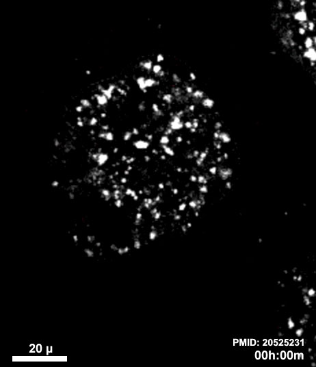
Mouse Embryo Lipid Droplets
|
| width=400px|height=425px|controller=true|autoplay=false</qt> | Live imaging of YFP-labelled Melanoblasts in Embryonic Mouse Skin (E14.5)
Cells that are actively migrating exhibit a characteristic spindle-like shape (red arrows), while dividing cells appear rounded (blue circles).
|
Reference: <pubmed>20067551</pubmed>| PMC2859249
MRC Human Genetics Unit, Institute of Genetics and Molecular Medicine, Western General Hospital, Edinburgh, UK Professor Ian J. Jackson, e-mail: Ian.Jackson@hgu.mrc.ac.uk
Re-use of this article is permitted in accordance with the Creative Commons Deed, Attribution 2.5, which does not permit commercial exploitation.