Mouse Melanoblast Migration Movie: Difference between revisions
mNo edit summary |
mNo edit summary |
||
| Line 2: | Line 2: | ||
{| | {| | ||
| <mediaplayer width='450' height='470' image="http://embryology.med.unsw.edu.au/embryology/images/9/93/Mouse-melanoblast_migration_icon.jpg">File:Mouse_melanoblast_migration.mp4</mediaplayer> | | <mediaplayer width='450' height='470' image="http://embryology.med.unsw.edu.au/embryology/images/9/93/Mouse-melanoblast_migration_icon.jpg">File:Mouse_melanoblast_migration.mp4</mediaplayer> | ||
| valign="top" | | | valign="top" | | ||
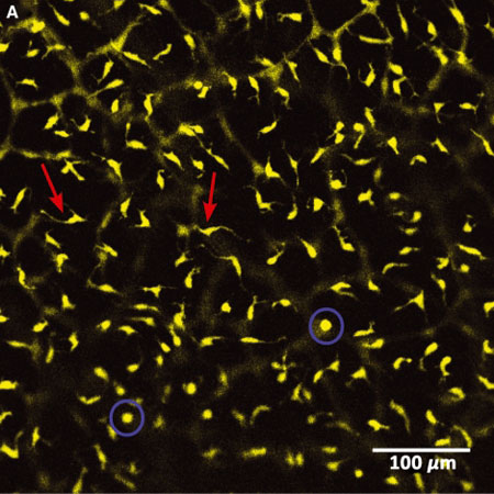
Live imaging of YFP-labelled Melanoblasts in Embryonic Mouse Skin (E14.5) | Live imaging of YFP-labelled Melanoblasts in Embryonic Mouse Skin (E14.5) | ||
Cells that are actively migrating exhibit a characteristic spindle-like shape (red arrows), while dividing cells appear rounded (blue circles). | Cells that are actively migrating exhibit a characteristic spindle-like shape (red arrows), while dividing cells appear rounded (blue circles). | ||
[[File:Mouse-melanoblast migration icon.jpg|thumb]] | |||
'''Links:''' [[Media:Mouse_melanoblast_migration.mp4|MP4 version]] | [[Neural Crest Development]] | [[Integumentary System Development]] | [[Mouse Development]] | '''Links:''' [[Media:Mouse_melanoblast_migration.mp4|MP4 version]] | [[Neural Crest Development]] | [[Integumentary System Development]] | [[Mouse Development]] | ||
Revision as of 11:51, 23 March 2014
| Embryology - 27 Apr 2024 |
|---|
| Google Translate - select your language from the list shown below (this will open a new external page) |
|
العربية | català | 中文 | 中國傳統的 | français | Deutsche | עִברִית | हिंदी | bahasa Indonesia | italiano | 日本語 | 한국어 | မြန်မာ | Pilipino | Polskie | português | ਪੰਜਾਬੀ ਦੇ | Română | русский | Español | Swahili | Svensk | ไทย | Türkçe | اردو | ייִדיש | Tiếng Việt These external translations are automated and may not be accurate. (More? About Translations) |
| <mediaplayer width='450' height='470' image="http://embryology.med.unsw.edu.au/embryology/images/9/93/Mouse-melanoblast_migration_icon.jpg">File:Mouse_melanoblast_migration.mp4</mediaplayer> |
Live imaging of YFP-labelled Melanoblasts in Embryonic Mouse Skin (E14.5) Cells that are actively migrating exhibit a characteristic spindle-like shape (red arrows), while dividing cells appear rounded (blue circles). Links: MP4 version | Neural Crest Development | Integumentary System Development | Mouse Development |
Reference
<pubmed>20067551</pubmed>| PMC2859249
Copyright
Re-use of this article is permitted in accordance with the Creative Commons Deed, Attribution 2.5, which does not permit commercial exploitation.
MRC Human Genetics Unit, Institute of Genetics and Molecular Medicine, Western General Hospital, Edinburgh, UK Professor Ian J. Jackson, e-mail: Ian.Jackson@hgu.mrc.ac.uk