Human Embryology and Morphology - Figures: Difference between revisions
mNo edit summary |
|||
| (4 intermediate revisions by the same user not shown) | |||
| Line 1: | Line 1: | ||
{{Header}} | |||
{{Keith1921 header}} | {{Keith1921 header}} | ||
| Line 167: | Line 168: | ||
<gallery> | <gallery> | ||
File:Keith1921 fig106.jpg| | File:Keith1921 fig106.jpg|Fig. 106. Cerebral Vesicle and the formation of the Corpus Striatum on its floor, during the 6th week. | ||
File:Keith1921 fig107.jpg| | File:Keith1921 fig107.jpg|Fig. 107. Expansion of the left Cerebral Vesicle as seen on its lateral aspect. | ||
File:Keith1921 fig108.jpg| | File:Keith1921 fig108.jpg|Fig. 108. Fore- and Mid-Brain at the 6th week of development. | ||
File:Keith1921 fig109.jpg| | File:Keith1921 fig109.jpg|Fig. 109. 3rd and lateral Ventricles of the Adult. | ||
File:Keith1921 fig110.jpg| | File:Keith1921 fig110.jpg|Fig. 110. Turtle's Cerebral Vesicle and Primitive Mammalian Cerebrum. | ||
File:Keith1921 fig111.jpg| | File:Keith1921 fig111.jpg|Fig. 111. Differentiation of the Pallial Wall of the Cerebral Vesicle. | ||
File:Keith1921 fig112.jpg| | File:Keith1921 fig112.jpg|Fig. 112. Left Hemisphere of the Brain of a primitive vertebrate brain anterior to the Lamina Terminalis. | ||
File:Keith1921 fig113.jpg| | File:Keith1921 fig113.jpg|Fig. 113. Coronal section of the right half of the Cerebral Vesicle of a Primitive Type of Mammal. | ||
File:Keith1921 fig114.jpg| | File:Keith1921 fig114.jpg|Fig. 114. Lateral Aspect of the Cerebrum of a Primitive Mammal. | ||
File:Keith1921 fig115.jpg| | File:Keith1921 fig115.jpg|Fig. 115. The Anterior, Hippocampal and Callosal Commissures. | ||
File:Keith1921 fig116.jpg| | File:Keith1921 fig116.jpg|Fig. 116. Mesial Aspect of the Brain of a Human Foetus in 4th month. | ||
File:Keith1921 fig117.jpg| | File:Keith1921 fig117.jpg|Fig. 117. Mesial aspect of the Cerebral Vesicle of a Foetus about 3 months old. | ||
File:Keith1921 fig118.jpg| | File:Keith1921 fig118.jpg|Fig. 118. Structures formed in the Lamina Terminalis and Primitive Callosal Gyrus. | ||
File:Keith1921 fig119.jpg| | File:Keith1921 fig119.jpg|Fig. 119. Lateral Aspect of the Cerebral Hemisphere at the end of the 2nd month | ||
File:Keith1921 fig120.jpg| | File:Keith1921 fig120.jpg|Fig. 120. The same Aspect during the 5th month. | ||
File:Keith1921 fig121.jpg| | File:Keith1921 fig121.jpg|Fig. 121. The same Aspect during the 7th month. | ||
File:Keith1921 fig122.jpg| | File:Keith1921 fig122.jpg|Fig. 122. Opercula and Fissure of Sylvius. | ||
File:Keith1921 fig123.jpg| | File:Keith1921 fig123.jpg|Fig. 123. The Island of Reil and Fissures on the Lateral Aspect of the Brain of a dog-like Ape. | ||
File:Keith1921 fig124.jpg| | File:Keith1921 fig124.jpg|Fig. 124. The more common condition of the Island of Reil in Anthropoids. | ||
File:Keith1921 fig125.jpg| | File:Keith1921 fig125.jpg|Fig. 125. The Fissures on the Lateral Aspect of a typical Mammalian Brain. | ||
File:Keith1921 fig126.jpg| | File:Keith1921 fig126.jpg|Fig. 126. The Lateral Aspect of the Occipital Lobe of a Human Brain | ||
File:Keith1921 fig127.jpg| | File:Keith1921 fig127.jpg|Fig. 127. The Mesial Aspect of the Occipital Lobe of a Human Brain | ||
File:Keith1921 fig128.jpg| | File:Keith1921 fig128.jpg|Fig. 128. Arteries of the Brain end of the first month. | ||
File:Keith1921 fig129.jpg| | File:Keith1921 fig129.jpg|Fig. 129. The Primitive Vein of the Head and its tributaries in the 6th week | ||
</gallery> | </gallery> | ||
| Line 197: | Line 198: | ||
<gallery> | <gallery> | ||
File:Keith1921 fig130.jpg| | File:Keith1921 fig130.jpg|Fig. 130. The Chondrocranium of a Shark laid open by a mesial sagittal section. | ||
File:Keith1921 fig131.jpg| | File:Keith1921 fig131.jpg| | ||
File:Keith1921 fig132.jpg| | File:Keith1921 fig132.jpg| | ||
| Line 662: | Line 663: | ||
{{Keith1921 footer}} | {{Keith1921 footer}} | ||
[[Category:Draft]] | |||
Latest revision as of 11:00, 18 November 2016
| Embryology - 27 Apr 2024 |
|---|
| Google Translate - select your language from the list shown below (this will open a new external page) |
|
العربية | català | 中文 | 中國傳統的 | français | Deutsche | עִברִית | हिंदी | bahasa Indonesia | italiano | 日本語 | 한국어 | မြန်မာ | Pilipino | Polskie | português | ਪੰਜਾਬੀ ਦੇ | Română | русский | Español | Swahili | Svensk | ไทย | Türkçe | اردو | ייִדיש | Tiếng Việt These external translations are automated and may not be accurate. (More? About Translations) |
Keith, A. Human Embryology And Morphology (1921) Longmans, Green & Co.:New York.
Human Embryology and Morphology: 1 Early Ovum and Embryo | 2 Connection between Foetus and Uterus | 3 Primitive Streak Notochord and Somites | 4 Age Changes | 5 Spinal Column and Back | 6 Body Segmentation | 7 Spinal Cord | 8 Mid- and Hind-Brains | 9 Fore-Brain | 10 Fore-Brain Cerebral Vesicles | 11 Cranium | 12 Face | 13 Teeth and Mastication | 14 Nasal and Olfactory | 15 Sense OF Sight | 16 Hearing | 17 Pharynx and Neck | 18 Tongue, Thyroid and Pharynx | 19 Organs of Digestion | 20 Circulatory System | 21 Circulatory System (continued) | 22 Respiratory System | 23 Urogenital System | 24 Urogenital System (Continued) | 25 Body Wall and Pelvic Floor | 26 Limb Buds | 27 Limbs | 28 Skin and Appendages | Figures
| Historic Disclaimer - information about historic embryology pages |
|---|
 |
Note some of these figures are the same as those used in the earlier 1902 first edition.
Early Changes in the Development of the Ovum and Embryo
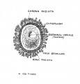
Fig. 1. Parts of a Mature Human Ovum
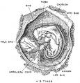
Fig. 2. Human Embryo and its Membranes at the end of the fifth week
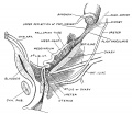
Fig. 3. Position of the Ovary and Fallopian Tube in the 5th month
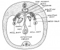
Fig. 4. Foetus at the beginning of the 3rd month
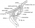
Fig. 5. Human Embryo and its Membranes at the end of the fifth week
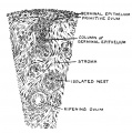
Fig. 6. Ovary of a fifth month Foetus
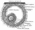
Fig. 7. Ripe Graafian Follicle at Puberty
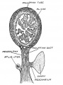
Fig. 8. Broad Ligament and Fallopian Tube
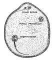
Fig. 9. Mature Ovum of Bat
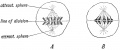
Fig. 10. Karyokinesis in a somatic cell
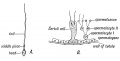
Fig. 11. Spermatozoon
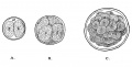
Fig. 12. Production of the Blastula or Morula from the Ovum
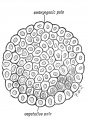
Fig. 13. Stage I. The Blastula
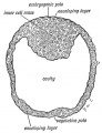
Fig. 14. The Blastocyst
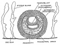
Fig. 15. Stage II. The Blasto-dermic Stage
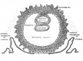
Fig. 16. Origin of the Primitive Coelom, the Mesoblast and Cavity of the Amnion
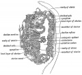
Fig. 17. Stage IV. Section through the bivesicular blastocyst embedded in the wall of the Uterus
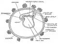
Fig. 18. Stage V. Diagrammatic Section of a human pregnancy towards the end of the 3rd week
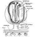
Fig. 19. Medullary folds and somites on the embryonic plate

Fig. 20. Human Embryo 2.5 mm long towards the end of the fourth week
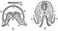
Fig. 21. Schematic Transverse Sections of two Human Embryos
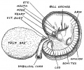
Fig. 22. Showing a human embryo 5 mm in length
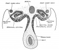
Fig. 23. Roof of the Coelomic cavity of a human embryo in the fifth week


The Manner in which a Connection is Established between the Foetus and Uterus
2 Connection between Foetus and Uterus
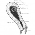
Fig. 24. Uterus showing the Embedded Ovum and the Decidua Three Parts
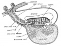
Fig. 25. The Yolk Sac and early vessels of the human embryo about the end of the 3rd week of development
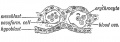
Fig. 26. Yolk Sac, blood vessels and nucleated red blood corpuscles in its mesoblastic layer
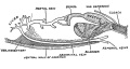
Fig. 27. The cloaca, bladder and abdominal vein of a frog
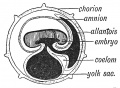
Fig. 28. The primitive form of the Allantois
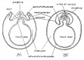
Fig. 29. Chorion and amnion arise in the chick embryo from folds of the somatopleure
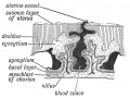
Fig. 30. Decidua Serotina (formed frcm the mucous membrane of uterus) and Chorion
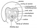
Fig. 31. Section across the Body-Stalk
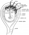
Fig. 32. Amnion, Chorion, and Decidua in the 3rd month and the Formation of the Placenta
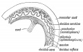
Fig. 33. Elements in the formation of the Placenta


The Primitive Streak, Notochord and Somites
3 Primitive Streak Notochord and Somites
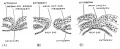
Fig. 34. Human embryonic plate measuring 1.5 mm in length
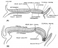
Fig. 35. Sections along the median line of two embryonic plates
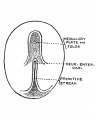
Fig. 36. Embryogenic area of an Embryonic plate viewed from above
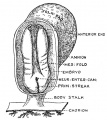
Fig. 37. The Medullary Plate and Primitive Streak on an Embryo towards the end of the 3rd week
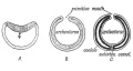
Fig. 38.Three stages in the early development of Amphioxus
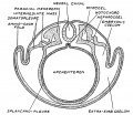
Fig. 39. Vertebrate embryo to show the parts of the mesoderm, of the coelom
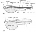
Fig. 40. Diagrammatic longitudinal section of a larval ganoid fish
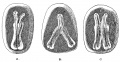
Fig. 41. Division of the Embryonic Plate forming imperfect twins


The Age Changes in the Embryo and Foetus
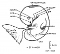
Fig. 42. Human Embryo 5 mm in length
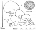
Fig. 43. Stages of growth from 3rd week to the end of the 8th
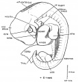
Fig. 44. Human Embryo 10.4 mm long and in the 6th week of development
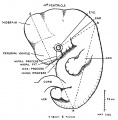
Fig. 45. Embryo 11 mm long
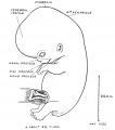
Fig. 46. Foetus 22 mm long


The Spinal Column and Back
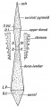
Fig. 47. Pyramids of the Spine
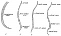
Fig. 48. Curves of the Spinal Column
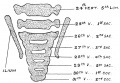
Fig. 49. Lumbo-sacral Region Spine Foetus end of the 2nd month
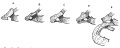
Fig. 50. Variation in costal element of the seventh Cervical Vertebra
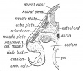
Fig. 51. Sclerotome, muscle plate and skin plate
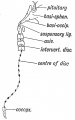
Fig. 52. Where remnants of the Notochord may occur in the Adult
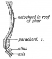
Fig. 53. The relationship of the Notochord to the basilar or parachordal cartilage

Fig. 54. The Morphological Parts of the first Cervical Vertebra
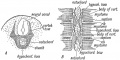
Fig. 55. The development of the Membranous Basis of a Vertebra
Fig. 56. Showing the Stages in the Development of a Vertebra
Fig. 57. The Order in which the Centres of Ossification appear in the Bodies
Fig. 58. A Diagrammatic Section of the Foetal Axis, Atlas, and Basi-occipital
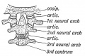
Fig. 59. The nature of the Atlanto-axio-occipital Articulations
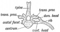
Fig. 60. The Bicipital Rib of a Lower Vertebrate (crocodile)
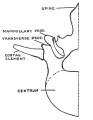
Fig. 61. Half of a first Lumbar Vertebra showing a separate costal element
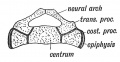
Fig. 62. A Section to show the Nature of the Elements composing the Sacrum
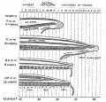
Fig. 63. A series of four figures showing the conlition of the liuman caudal or coccygeal region


The Segmentation of the Body
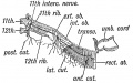
Fig. 64. Some of the structures derived from the 11th Dorsal Segment of the Right Side.
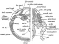
Fig. 65 Transverse Section showing the Elements of the 1st Lumbar Segment in the Adult
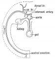
Fig. 66. The distribution of a typical Segmental Artery.
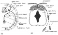
Fig. 67. Nerve System of the 11th Dorsal Segment.
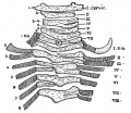
Fig. 68. Cervical and dorsal parts of the Spine of a Human Foetus showing irregularities of segmentation.


Central Nervous System — Differentiation of the Spinal Cord
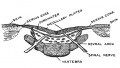
Fig. 69. Back of an Anencephalic Child in which the medullary plates were exposed on both head and spine.
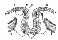
Fig. 70. Ectodermal cells of the Medullary Plates are differentiated into nerve cells or neuroblasts and supporting cells or spongioblasts.
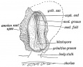
Fig. 71. Medullary Folds uniting to form the Neural Tube in a Human Embryo in the 3rd week of development.

Fig. 72. Four Primary Divisions of the Neural Tube.
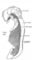
Fig. 73. Lateral View of the Central Nerve System of a Human Embryo of the 4th week
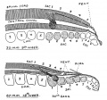
Fig. 73, A. Showing the differentiation of the terminal part of the neural tube into the coccygeal thread and fllum terminale.
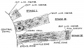
Fig. 74. Three stages in the early differentiation of the wall of the spinal neural tube.
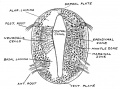
Fig. 75. Developing Spinal Cord at the beginning of the 5th week.
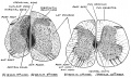
Fig. 76. Progressive differentiation of the spinal cord during the second and third months of development.
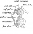
Fig. 77. Section of the developing Spinal Cord
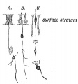
Fig. 78. Transformation of Cells of the Ectoderm to Sense Epithelium, Nerve Cells and Supporting Cells
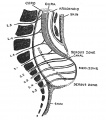
Fig. 79. Lumbar Region to show the arrangement of parts in a typical case of cystic spina bifida.


The Mid- and Hind-Brains
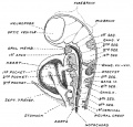
Fig. 80. Tubular form, the neuromeres and relations of the Mid- and Hind-Brain in a Human Embryo 18 body somites
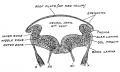
Fig. 81. Hind-Brain of a Human Embryo in the 6th week.
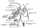
Fig. 82. Hind-Brain to show the grouping of cranial nerves and their nuclei.
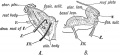
Fig. 83. Foetal Medulla and the Alar and Basal Laminae of the Hind-Brain
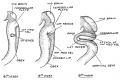
Fig. 84. Origin of the Inferior Medullary Velum from the roof plate of the Hind-Brain.
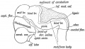
Fig. 85. Lateral View of the Cephalic Part of the Neural Tube in a 5th week Human Embryo.
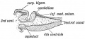
Fig. 86. Median Section of the Cerebellum and 4th Ventricle of a Frog.
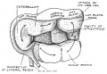
Fig. 87. The Human Cerebellum at the end of the 2nd month of development.
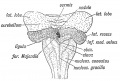
Fig. 88. Cerebellum and Attachments of the Inferior Medullary Velum at 3rd month
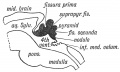
Fig. 89. Cerebellum of a Human Foetus early in the 4th month
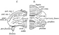
Fig. 90. Left and Right half of the Cerebellum of a Foetus of 5 months
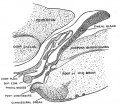
Fig. 91. Anterior part of the Roof of the Mid-Brain of a Cat, to show the subcommissural organ.
Fig. 92. Diagrammatic Section across the posterior region of the Head of Ammoecetes
Fig. 93. The Nerves and Ganglia of the Mid- and Hind-Brain of an Embryo at the end of the 6th week


The Fore-Brain or Prosencephalon
Fig. 94. Brain of a Larval Fish primary form and relations of the fore-brain.
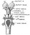
Fig. 95. The Brain of the Lamprey from above.
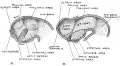
Fig. 96. Fore-Brain at the beginning and end of the 4th week.
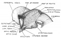
Fig. 97. The Thalamencephalon towards the end of the 6th week.
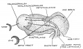
Fig. 98. Embryonic Fore-Brain various parts linked to sensory tracts.
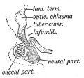
Fig. 99. Pituitary Body of a Human Foetus in the 5th month.
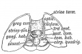
Fig. 100. Pineal Body and its commissure and ganglion.
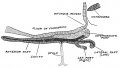
Fig. 101. Pituitary Body of a Pup Dog-Fish.
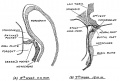
Fig. 102. Development of the Pituitary.
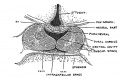
Fig. 103. Pituitary Body of a Human Foetus at the beginning of the 4th month.
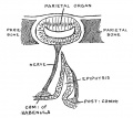
Fig. 104. The Pineal Gland and Sense Organ in a Lizard.
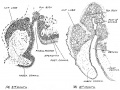
Fig. 105. Showing stages of development of the Pineal Body in the roof of the Fore-Brain


The Fore-Brain or Prosencephalon (continued) Cerebral Vesicles
10 Fore-Brain Cerebral Vesicles
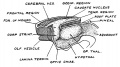
Fig. 106. Cerebral Vesicle and the formation of the Corpus Striatum on its floor, during the 6th week.
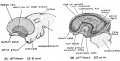
Fig. 107. Expansion of the left Cerebral Vesicle as seen on its lateral aspect.
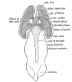
Fig. 108. Fore- and Mid-Brain at the 6th week of development.
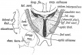
Fig. 109. 3rd and lateral Ventricles of the Adult.
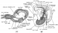
Fig. 110. Turtle's Cerebral Vesicle and Primitive Mammalian Cerebrum.
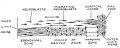
Fig. 111. Differentiation of the Pallial Wall of the Cerebral Vesicle.
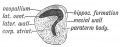
Fig. 112. Left Hemisphere of the Brain of a primitive vertebrate brain anterior to the Lamina Terminalis.
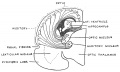
Fig. 113. Coronal section of the right half of the Cerebral Vesicle of a Primitive Type of Mammal.
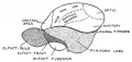
Fig. 114. Lateral Aspect of the Cerebrum of a Primitive Mammal.
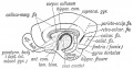
Fig. 115. The Anterior, Hippocampal and Callosal Commissures.
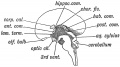
Fig. 116. Mesial Aspect of the Brain of a Human Foetus in 4th month.
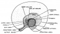
Fig. 117. Mesial aspect of the Cerebral Vesicle of a Foetus about 3 months old.
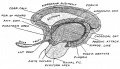
Fig. 118. Structures formed in the Lamina Terminalis and Primitive Callosal Gyrus.
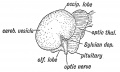
Fig. 119. Lateral Aspect of the Cerebral Hemisphere at the end of the 2nd month
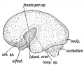
Fig. 120. The same Aspect during the 5th month.
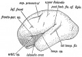
Fig. 121. The same Aspect during the 7th month.
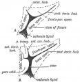
Fig. 122. Opercula and Fissure of Sylvius.
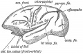
Fig. 123. The Island of Reil and Fissures on the Lateral Aspect of the Brain of a dog-like Ape.
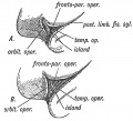
Fig. 124. The more common condition of the Island of Reil in Anthropoids.
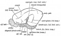
Fig. 125. The Fissures on the Lateral Aspect of a typical Mammalian Brain.
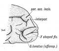
Fig. 126. The Lateral Aspect of the Occipital Lobe of a Human Brain
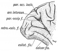
Fig. 127. The Mesial Aspect of the Occipital Lobe of a Human Brain
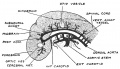
Fig. 128. Arteries of the Brain end of the first month.
Fig. 129. The Primitive Vein of the Head and its tributaries in the 6th week


The Cranium
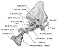
Fig. 130. The Chondrocranium of a Shark laid open by a mesial sagittal section.
- Keith1921 fig132.jpg
- Keith1921 fig133.jpg
- Keith1921 fig134.jpg
- Keith1921 fig135.jpg
- Keith1921 fig136.jpg
- Keith1921 fig137.jpg
- Keith1921 fig138.jpg
- Keith1921 fig139.jpg
- Keith1921 fig140.jpg
- Keith1921 fig141.jpg
- Keith1921 fig142.jpg
- Keith1921 fig143.jpg
- Keith1921 fig144.jpg
- Keith1921 fig145.jpg
- Keith1921 fig146.jpg
- Keith1921 fig147.jpg
- Keith1921 fig148.jpg
- Keith1921 fig149.jpg
- Keith1921 fig150.jpg


Development of the Face
- Keith1921 fig151.jpg
- Keith1921 fig152.jpg
- Keith1921 fig153.jpg
- Keith1921 fig154.jpg
- Keith1921 fig155.jpg
- Keith1921 fig156.jpg
- Keith1921 fig157.jpg
- Keith1921 fig158.jpg
- Keith1921 fig159.jpg
- Keith1921 fig160.jpg
- Keith1921 fig161.jpg
- Keith1921 fig162.jpg
- Keith1921 fig163.jpg
- Keith1921 fig164.jpg
- Keith1921 fig165.jpg
- Keith1921 fig166.jpg
- Keith1921 fig167.jpg
- Keith1921 fig168.jpg
- Keith1921 fig169.jpg
- Keith1921 fig170.jpg
- Keith1921 fig171.jpg
- Keith1921 fig172.jpg
- Keith1921 fig173.jpg
- Keith1921 fig174.jpg
- Keith1921 fig175.jpg
- Keith1921 fig176.jpg
- Keith1921 fig177.jpg
- Keith1921 fig178.jpg
- Keith1921 fig179.jpg
- Keith1921 fig180.jpg
- Keith1921 fig181.jpg
- Keith1921 fig182.jpg
- Keith1921 fig183.jpg
The Teeth and Apparatus of Mastication
- Keith1921 fig184.jpg
- Keith1921 fig185.jpg
- Keith1921 fig186.jpg
- Keith1921 fig187.jpg
- Keith1921 fig188.jpg
- Keith1921 fig189.jpg
- Keith1921 fig190.jpg
- Keith1921 fig191.jpg
The Nasal Cavities and Olfactory Structures
- Keith1921 fig192.jpg
- Keith1921 fig193.jpg
- Keith1921 fig194.jpg
- Keith1921 fig195.jpg
- Keith1921 fig196.jpg
- Keith1921 fig197.jpg
- Keith1921 fig198.jpg
- Keith1921 fig199.jpg
- Keith1921 fig200.jpg
- Keith1921 fig201.jpg
- Keith1921 fig202.jpg
Development of the Structures concerned in the Sense of Sight
- Keith1921 fig203.jpg
- Keith1921 fig204.jpg
- Keith1921 fig205.jpg
- Keith1921 fig206.jpg
- Keith1921 fig207.jpg
- Keith1921 fig208.jpg
- Keith1921 fig209.jpg
- Keith1921 fig210.jpg
- Keith1921 fig211.jpg
- Keith1921 fig212.jpg
- Keith1921 fig213.jpg
- Keith1921 fig214.jpg
- Keith1921 fig215.jpg
- Keith1921 fig216.jpg
- Keith1921 fig217.jpg
- Keith1921 fig218.jpg
- Keith1921 fig219.jpg
- Keith1921 fig220.jpg
- Keith1921 fig221.jpg
- Keith1921 fig222.jpg
- Keith1921 fig223.jpg
- Keith1921 fig224.jpg
- Keith1921 fig225.jpg
- Keith1921 fig226.jpg
- Keith1921 fig227.jpg
The Organ of Hearing
- Keith1921 fig228.jpg
- Keith1921 fig229.jpg
- Keith1921 fig230.jpg
- Keith1921 fig231.jpg
- Keith1921 fig232.jpg
- Keith1921 fig233.jpg
- Keith1921 fig234.jpg
- Keith1921 fig235.jpg
- Keith1921 fig236.jpg
- Keith1921 fig237.jpg
- Keith1921 fig238.jpg
- Keith1921 fig239.jpg
- Keith1921 fig240.jpg
- Keith1921 fig241.jpg
- Keith1921 fig242.jpg
- Keith1921 fig243.jpg
- Keith1921 fig244.jpg
- Keith1921 fig245.jpg
- Keith1921 fig246.jpg
- Keith1921 fig247.jpg
Pharynx and Neck
- Keith1921 fig248.jpg
- Keith1921 fig249.jpg
- Keith1921 fig250.jpg
- Keith1921 fig251.jpg
- Keith1921 fig252.jpg
- Keith1921 fig253.jpg
- Keith1921 fig254.jpg
- Keith1921 fig255.jpg
- Keith1921 fig256.jpg
- Keith1921 fig257.jpg
- Keith1921 fig258.jpg
- Keith1921 fig259.jpg
- Keith1921 fig260.jpg
- Keith1921 fig261.jpg
Tongue, Thyroid and Structures developed from the Walls of the Primitive Pharynx
18 Tongue, Thyroid and Pharynx
- Keith1921 fig262.jpg
- Keith1921 fig263.jpg
- Keith1921 fig264.jpg
- Keith1921 fig265.jpg
- Keith1921 fig266.jpg
- Keith1921 fig267.jpg
- Keith1921 fig268.jpg
- Keith1921 fig269.jpg
- Keith1921 fig270.jpg
- Keith1921 fig271.jpg
- Keith1921 fig272.jpg
Organs of Digestion
- Keith1921 fig273.jpg
- Keith1921 fig274.jpg
- Keith1921 fig275.jpg
- Keith1921 fig276.jpg
- Keith1921 fig277.jpg
- Keith1921 fig278.jpg
- Keith1921 fig279.jpg
- Keith1921 fig280.jpg
- Keith1921 fig281.jpg
- Keith1921 fig282.jpg
- Keith1921 fig283.jpg
- Keith1921 fig284.jpg
- Keith1921 fig285.jpg
- Keith1921 fig286.jpg
- Keith1921 fig287.jpg
- Keith1921 fig288.jpg
- Keith1921 fig289.jpg
- Keith1921 fig290.jpg
- Keith1921 fig291.jpg
- Keith1921 fig292.jpg
- Keith1921 fig293.jpg
- Keith1921 fig294.jpg
- Keith1921 fig295.jpg
- Keith1921 fig296.jpg
- Keith1921 fig297.jpg
- Keith1921 fig298.jpg
- Keith1921 fig299.jpg
- Keith1921 fig300.jpg
- Keith1921 fig301.jpg
- Keith1921 fig302.jpg
- Keith1921 fig303.jpg
- Keith1921 fig304.jpg
- Keith1921 fig305.jpg
- Keith1921 fig306.jpg
- Keith1921 fig307.jpg
- Keith1921 fig308.jpg
- Keith1921 fig309.jpg
Circulatory System
- Keith1921 fig312.jpg
- Keith1921 fig313.jpg
- Keith1921 fig314.jpg
- Keith1921 fig315.jpg
- Keith1921 fig316.jpg
- Keith1921 fig317.jpg
- Keith1921 fig318.jpg
- Keith1921 fig319.jpg
- Keith1921 fig320.jpg
- Keith1921 fig321.jpg
- Keith1921 fig322.jpg
- Keith1921 fig323.jpg
- Keith1921 fig324.jpg
- Keith1921 fig325.jpg
- Keith1921 fig326.jpg
- Keith1921 fig327.jpg
- Keith1921 fig328.jpg
- Keith1921 fig329.jpg
- Keith1921 fig330.jpg
- Keith1921 fig331.jpg
- Keith1921 fig332.jpg
- Keith1921 fig333.jpg
- Keith1921 fig334.jpg
- Keith1921 fig335.jpg
- Keith1921 fig336.jpg
- Keith1921 fig337.jpg
- Keith1921 fig338.jpg
- Keith1921 fig339.jpg
- Keith1921 fig340.jpg
- Keith1921 fig341.jpg
- Keith1921 fig342.jpg
- Keith1921 fig343.jpg
- Keith1921 fig344.jpg
- Keith1921 fig345.jpg
- Keith1921 fig346.jpg
- Keith1921 fig347.jpg
- Keith1921 fig348.jpg
- Keith1921 fig349.jpg
Circulatory System (continued)
21 Circulatory System (continued)
- Keith1921 fig350.jpg
- Keith1921 fig351.jpg
- Keith1921 fig352.jpg
- Keith1921 fig353.jpg
- Keith1921 fig354.jpg
- Keith1921 fig355.jpg
- Keith1921 fig356.jpg
- Keith1921 fig357.jpg
- Keith1921 fig358.jpg
- Keith1921 fig359.jpg
Respiratory System
- Keith1921 fig360.jpg
- Keith1921 fig361.jpg
- Keith1921 fig362.jpg
- Keith1921 fig363.jpg
- Keith1921 fig364.jpg
- Keith1921 fig365.jpg
- Keith1921 fig366.jpg
- Keith1921 fig367.jpg
- Keith1921 fig368.jpg
- Keith1921 fig369.jpg
- Keith1921 fig370.jpg
- Keith1921 fig371.jpg
- Keith1921 fig372.jpg
- Keith1921 fig373.jpg
- Keith1921 fig374.jpg
- Keith1921 fig375.jpg
- Keith1921 fig376.jpg
- Keith1921 fig377.jpg
- Keith1921 fig378.jpg
- Keith1921 fig379.jpg
Urogenital System
- Keith1921 fig380.jpg
- Keith1921 fig381.jpg
- Keith1921 fig382.jpg
- Keith1921 fig383.jpg
- Keith1921 fig384.jpg
- Keith1921 fig385.jpg
- Keith1921 fig386.jpg
- Keith1921 fig387.jpg
- Keith1921 fig388.jpg
- Keith1921 fig389.jpg
- Keith1921 fig390.jpg
- Keith1921 fig391.jpg
- Keith1921 fig392.jpg
- Keith1921 fig393.jpg
- Keith1921 fig394.jpg
- Keith1921 fig395.jpg
- Keith1921 fig396.jpg
- Keith1921 fig397.jpg
- Keith1921 fig398.jpg
- Keith1921 fig399.jpg
- Keith1921 fig400.jpg
- Keith1921 fig401.jpg
- Keith1921 fig402.jpg
Urogenital System (continued)
24 Urogenital System (Continued)
- Keith1921 fig403.jpg
- Keith1921 fig404.jpg
- Keith1921 fig405.jpg
- Keith1921 fig406.jpg
- Keith1921 fig407.jpg
- Keith1921 fig408.jpg
- Keith1921 fig409.jpg
- Keith1921 fig410.jpg
- Keith1921 fig411.jpg
- Keith1921 fig412.jpg
- Keith1921 fig413.jpg
- Keith1921 fig414.jpg
- Keith1921 fig415.jpg
- Keith1921 fig416.jpg
- Keith1921 fig417.jpg
- Keith1921 fig418.jpg
- Keith1921 fig419.jpg
- Keith1921 fig420.jpg
- Keith1921 fig421.jpg
- Keith1921 fig422.jpg
- Keith1921 fig423.jpg
- Keith1921 fig424.jpg
- Keith1921 fig425.jpg
- Keith1921 fig426.jpg
- Keith1921 fig427.jpg
Body Wall and Pelvic Floor
- Keith1921 fig428.jpg
- Keith1921 fig429.jpg
- Keith1921 fig430.jpg
- Keith1921 fig431.jpg
- Keith1921 fig432.jpg
- Keith1921 fig433.jpg
- Keith1921 fig434.jpg
- Keith1921 fig435.jpg
- Keith1921 fig436.jpg
- Keith1921 fig437.jpg
- Keith1921 fig438.jpg
- Keith1921 fig439.jpg
- Keith1921 fig440.jpg
- Keith1921 fig441.jpg
- Keith1921 fig442.jpg
- Keith1921 fig443.jpg
- Keith1921 fig444.jpg
- Keith1921 fig445.jpg
- Keith1921 fig446.jpg
Development and Differentiation of the Limb Buds
- Keith1921 fig447.jpg
- Keith1921 fig448.jpg
- Keith1921 fig449.jpg
- Keith1921 fig450.jpg
- Keith1921 fig451.jpg
- Keith1921 fig452.jpg
- Keith1921 fig453.jpg
- Keith1921 fig454.jpg
- Keith1921 fig455.jpg
- Keith1921 fig456.jpg
- Keith1921 fig457.jpg
- Keith1921 fig458.jpg
- Keith1921 fig459.jpg
- Keith1921 fig460.jpg
- Keith1921 fig461.jpg
- Keith1921 fig462.jpg
Morphology of the Limbs
- Keith1921 fig463.jpg
- Keith1921 fig464.jpg
- Keith1921 fig465.jpg
- Keith1921 fig466.jpg
- Keith1921 fig467.jpg
- Keith1921 fig468.jpg
- Keith1921 fig469.jpg
- Keith1921 fig470.jpg
- Keith1921 fig471.jpg
- Keith1921 fig472.jpg
- Keith1921 fig473.jpg
- Keith1921 fig474.jpg
- Keith1921 fig475.jpg
- Keith1921 fig476.jpg
- Keith1921 fig477.jpg
- Keith1921 fig478.jpg
- Keith1921 fig479.jpg
Skin and its Appendages
- Keith1921 fig480.jpg
- Keith1921 fig481.jpg
- Keith1921 fig482.jpg
- Keith1921 fig483.jpg
- Keith1921 fig484.jpg
- Keith1921 fig485.jpg
- Keith1921 fig486.jpg
- Keith1921 fig487.jpg
- Keith1921 fig488.jpg
- Keith1921 fig489.jpg
Fig. 490. Breast Arrangement of its Capsule and Lymphatics.

| Historic Disclaimer - information about historic embryology pages |
|---|
|
Human Embryology and Morphology: 1 Early Ovum and Embryo | 2 Connection between Foetus and Uterus | 3 Primitive Streak Notochord and Somites | 4 Age Changes | 5 Spinal Column and Back | 6 Body Segmentation | 7 Spinal Cord | 8 Mid- and Hind-Brains | 9 Fore-Brain | 10 Fore-Brain Cerebral Vesicles | 11 Cranium | 12 Face | 13 Teeth and Mastication | 14 Nasal and Olfactory | 15 Sense OF Sight | 16 Hearing | 17 Pharynx and Neck | 18 Tongue, Thyroid and Pharynx | 19 Organs of Digestion | 20 Circulatory System | 21 Circulatory System (continued) | 22 Respiratory System | 23 Urogenital System | 24 Urogenital System (Continued) | 25 Body Wall and Pelvic Floor | 26 Limb Buds | 27 Limbs | 28 Skin and Appendages | Figures