Human Embryology and Morphology - Figures: Difference between revisions
| Line 5: | Line 5: | ||
<gallery> | <gallery> | ||
File:Keith1921 fig001.jpg|Fig. 1. | File:Keith1921 fig001.jpg|Fig. 1. Parts of a Mature Human Ovum | ||
File:Keith1921 fig002.jpg|Fig. 2. Human Embryo and its Membranes at the end of the fifth week | File:Keith1921 fig002.jpg|Fig. 2. Human Embryo and its Membranes at the end of the fifth week | ||
File:Keith1921 fig003.jpg|Fig. 3. | File:Keith1921 fig003.jpg|Fig. 3. Position of the Ovary and Fallopian Tube in the 5th month | ||
File:Keith1921 fig004.jpg|Fig. 4. | File:Keith1921 fig004.jpg|Fig. 4. Foetus at the beginning of the 3rd month | ||
File:Keith1921 fig005.jpg|Fig. 5. Human Embryo and its Membranes at the end of the fifth week | File:Keith1921 fig005.jpg|Fig. 5. Human Embryo and its Membranes at the end of the fifth week | ||
File:Keith1921 fig006.jpg|Fig. 6. | File:Keith1921 fig006.jpg|Fig. 6. Ovary of a fifth month Foetus | ||
File:Keith1921 fig007.jpg|Fig. 7. Ripe Graafian Follicle at Puberty | File:Keith1921 fig007.jpg|Fig. 7. Ripe Graafian Follicle at Puberty | ||
File:Keith1921 fig008.jpg|Fig. 8. | File:Keith1921 fig008.jpg|Fig. 8. Broad Ligament and Fallopian Tube | ||
File:Keith1921 fig009.jpg|Fig. 9. Mature Ovum of Bat | File:Keith1921 fig009.jpg|Fig. 9. Mature Ovum of Bat | ||
File:Keith1921 fig010.jpg|Fig. 10. | File:Keith1921 fig010.jpg|Fig. 10. Karyokinesis in a somatic cell | ||
File:Keith1921 fig011.jpg|Fig. 11. Spermatozoon | File:Keith1921 fig011.jpg|Fig. 11. Spermatozoon | ||
File:Keith1921 fig012.jpg|Fig. 12. | File:Keith1921 fig012.jpg|Fig. 12. Production of the Blastula or Morula from the Ovum | ||
File:Keith1921 fig013.jpg|Fig. 13. Stage I. The Blastula | File:Keith1921 fig013.jpg|Fig. 13. Stage I. The Blastula | ||
File:Keith1921 fig014.jpg|Fig. 14. The Blastocyst | File:Keith1921 fig014.jpg|Fig. 14. The Blastocyst | ||
File:Keith1921 fig015.jpg|Fig. 15. Stage II. The Blasto-dermic Stage | File:Keith1921 fig015.jpg|Fig. 15. Stage II. The Blasto-dermic Stage | ||
File:Keith1921 fig016.jpg|Fig. 16. | File:Keith1921 fig016.jpg|Fig. 16. Origin of the Primitive Coelom, the Mesoblast and Cavity of the Amnion | ||
File:Keith1921 fig017.jpg|Fig. 17. Stage IV. Section through the bivesicular blastocyst embedded in the wall of the Uterus | File:Keith1921 fig017.jpg|Fig. 17. Stage IV. Section through the bivesicular blastocyst embedded in the wall of the Uterus | ||
File:Keith1921 fig018.jpg|Fig. 18. Stage V. Diagrammatic Section of a human pregnancy towards the end of the 3rd week | File:Keith1921 fig018.jpg|Fig. 18. Stage V. Diagrammatic Section of a human pregnancy towards the end of the 3rd week | ||
File:Keith1921 fig019.jpg|Fig. 19. | File:Keith1921 fig019.jpg|Fig. 19. Medullary folds and somites on the embryonic plate | ||
File:Keith1921 fig020.jpg|Fig. 20. Human Embryo 2.5 mm long towards the end of the fourth week | File:Keith1921 fig020.jpg|Fig. 20. Human Embryo 2.5 mm long towards the end of the fourth week | ||
File:Keith1921 fig021.jpg| | |||
File:Keith1921 fig022.jpg| | |||
File:Keith1921 fig023.jpg| | |||
</gallery> | </gallery> | ||
Revision as of 05:52, 26 December 2014
Keith, A. Human Embryology And Morphology (1921) Longmans, Green & Co.:New York.
Human Embryology and Morphology: 1 Early Ovum and Embryo | 2 Connection between Foetus and Uterus | 3 Primitive Streak Notochord and Somites | 4 Age Changes | 5 Spinal Column and Back | 6 Body Segmentation | 7 Spinal Cord | 8 Mid- and Hind-Brains | 9 Fore-Brain | 10 Fore-Brain Cerebral Vesicles | 11 Cranium | 12 Face | 13 Teeth and Mastication | 14 Nasal and Olfactory | 15 Sense OF Sight | 16 Hearing | 17 Pharynx and Neck | 18 Tongue, Thyroid and Pharynx | 19 Organs of Digestion | 20 Circulatory System | 21 Circulatory System (continued) | 22 Respiratory System | 23 Urogenital System | 24 Urogenital System (Continued) | 25 Body Wall and Pelvic Floor | 26 Limb Buds | 27 Limbs | 28 Skin and Appendages | Figures
| Historic Disclaimer - information about historic embryology pages |
|---|
 |
Early Changes in the Development of the Ovum and Embryo
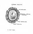
Fig. 1. Parts of a Mature Human Ovum
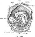
Fig. 2. Human Embryo and its Membranes at the end of the fifth week
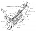
Fig. 3. Position of the Ovary and Fallopian Tube in the 5th month
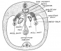
Fig. 4. Foetus at the beginning of the 3rd month
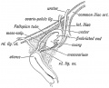
Fig. 5. Human Embryo and its Membranes at the end of the fifth week
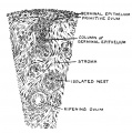
Fig. 6. Ovary of a fifth month Foetus
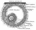
Fig. 7. Ripe Graafian Follicle at Puberty
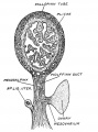
Fig. 8. Broad Ligament and Fallopian Tube
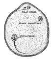
Fig. 9. Mature Ovum of Bat
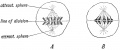
Fig. 10. Karyokinesis in a somatic cell
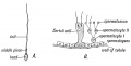
Fig. 11. Spermatozoon
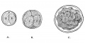
Fig. 12. Production of the Blastula or Morula from the Ovum
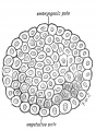
Fig. 13. Stage I. The Blastula
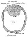
Fig. 14. The Blastocyst
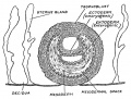
Fig. 15. Stage II. The Blasto-dermic Stage
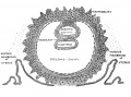
Fig. 16. Origin of the Primitive Coelom, the Mesoblast and Cavity of the Amnion
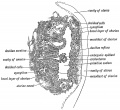
Fig. 17. Stage IV. Section through the bivesicular blastocyst embedded in the wall of the Uterus
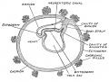
Fig. 18. Stage V. Diagrammatic Section of a human pregnancy towards the end of the 3rd week
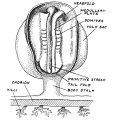
Fig. 19. Medullary folds and somites on the embryonic plate

Fig. 20. Human Embryo 2.5 mm long towards the end of the fourth week


The Manner in which a Connection is Established between the Foetus and Uterus
2 Connection between Foetus and Uterus
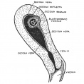
Fig. 24. Uterus showing the Embedded Ovum and the Decidua Three Parts
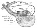
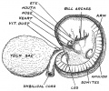
Fig. 25. The Yolk Sac and early vessels of the human embryo about the end of the 3rd week of development
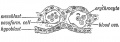
Fig. 26. Yolk Sac, blood vessels and nucleated red blood corpuscles in its mesoblastic layer
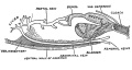
Fig. 27. The cloaca, bladder and abdominal vein of a frog
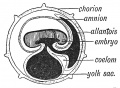
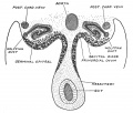
Fig. 28. The primitive form of the Allantois
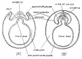
Fig. 29. Chorion and amnion arise in the chick embryo from folds of the somatopleure
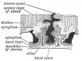
Fig. 30. Decidua Serotina (formed frcm the mucous membrane of uterus) and Chorion
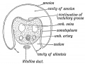
Fig. 31. Section across the Body-Stalk
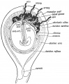
Fig. 32. Amnion, Chorion, and Decidua in the 3rd month and the Formation of the Placenta
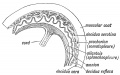
Fig. 33. Elements in the formation of the Placenta


The Primitive Streak, Notochord and Somites
3 Primitive Streak Notochord and Somites
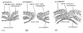
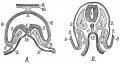
Fig. 34. Human embryonic plate measuring 1.5 mm in length
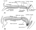
Fig. 35. Sections along the median line of two embryonic plates
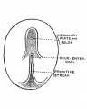
Fig. 36. Embryogenic area of an Embryonic plate viewed from above
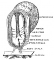
Fig. 37. The Medullary Plate and Primitive Streak on an Embryo towards the end of the 3rd week
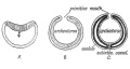
Fig. 38.Three stages in the early development of Amphioxus
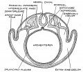
Fig. 39. Vertebrate embryo to show the parts of the mesoderm, of the coelom
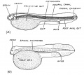
Fig. 40. Diagrammatic longitudinal section of a larval ganoid fish
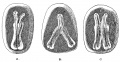
Fig. 41. Division of the Embryonic Plate forming imperfect twins


The Age Changes in the Embryo and Foetus
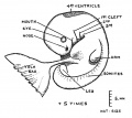
Fig. 42. Human Embryo 5 mm in length
Fig. 43. Stages of growth from 3rd week to the end of the 8th
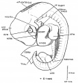
Fig. 44. Human Embryo 10.4 mm long and in the 6th week of development
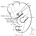
Fig. 45. Embryo 11 mm long
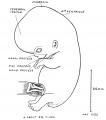
Fig. 46. Foetus 22 mm long


The Spinal Column and Back
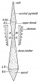
Fig. 47. Pyramids of the Spine
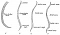
Fig. 48. Curves of the Spinal Column
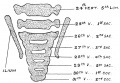
Fig. 49. Lumbo-sacral Region Spine Foetus end of the 2nd month
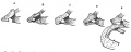
Fig. 50. Variation in costal element of the seventh Cervical Vertebra
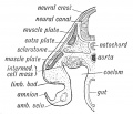
Fig. 51. Sclerotome, muscle plate and skin plate
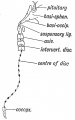
Fig. 52. Where remnants of the Notochord may occur in the Adult
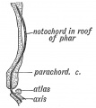
Fig. 53. The relationship of the Notochord to the basilar or parachordal cartilage

Fig. 54. The Morphological Parts of the first Cervical Vertebra
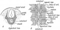
Fig. 55. The development of the Membranous Basis of a Vertebra
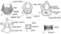
Fig. 56. Showing the Stages in the Development of a Vertebra
Fig. 57. The Order in which the Centres of Ossification appear in the Bodies
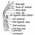
Fig. 58. A Diagrammatic Section of the Foetal Axis, Atlas, and Basi-occipital
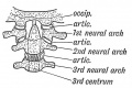
Fig. 59. The nature of the Atlanto-axio-occipital Articulations
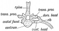
Fig. 60. The Bicipital Rib of a Lower Vertebrate (crocodile)
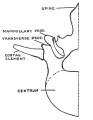
Fig. 61. Half of a first Lumbar Vertebra showing a separate costal element
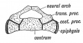
Fig. 62. A Section to show the Nature of the Elements composing the Sacrum
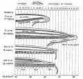
Fig. 63. A series of four figures showing the conlition of the liuman caudal or coccygeal region


The Segmentation of the Body
Central Nervous System — Differentiation of the Spinal Cord
The Mid- and Hind-Brains
The Fore-Brain or Prosencephalon
The Fore-Brain or Prosencephalon (continued). Cerebral Vesicles
10 Fore-Brain Cerebral Vesicles
The Cranium
Development of the Face
- Keith1921 fig151.jpg
Fig. 151.
The Teeth and Apparatus of Mastication
- Keith1921 fig184.jpg
Fig. 184.
The Nasal Cavities and Olfactory Structures
Development of the Structures concerned in the Sense OF Sight
The Organ of Hearing
Pharynx and Neck
Tongue, Thyroid and Structures developed from the Walls of the Primitive Pharynx
18 Tongue, Thyroid and Pharynx
Organs of Digestion
Circulatory System
Circulatory System (continued)
21 Circulatory System (continued)
Respiratory System
Urogenital System
Urogenital System (Continued)
24 Urogenital System (Continued)
Body Wall and Pelvic Floor
Development and Differentiation of the Limb Buds
Morphology of the Limbs
Skin and its Appendages
| Historic Disclaimer - information about historic embryology pages |
|---|
|
Human Embryology and Morphology: 1 Early Ovum and Embryo | 2 Connection between Foetus and Uterus | 3 Primitive Streak Notochord and Somites | 4 Age Changes | 5 Spinal Column and Back | 6 Body Segmentation | 7 Spinal Cord | 8 Mid- and Hind-Brains | 9 Fore-Brain | 10 Fore-Brain Cerebral Vesicles | 11 Cranium | 12 Face | 13 Teeth and Mastication | 14 Nasal and Olfactory | 15 Sense OF Sight | 16 Hearing | 17 Pharynx and Neck | 18 Tongue, Thyroid and Pharynx | 19 Organs of Digestion | 20 Circulatory System | 21 Circulatory System (continued) | 22 Respiratory System | 23 Urogenital System | 24 Urogenital System (Continued) | 25 Body Wall and Pelvic Floor | 26 Limb Buds | 27 Limbs | 28 Skin and Appendages | Figures