Human Embryo SEM: Difference between revisions
mNo edit summary |
m (→Introduction) |
||
| Line 2: | Line 2: | ||
==Introduction== | ==Introduction== | ||
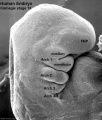
The following images are excerpts from a collaborative eBook currently in preparation. | The following images are image excerpts from a collaborative eBook currently in preparation. The images include both brightfieqld and scanning EM images of teh early human embryo between stage 7 to 14 (week 3-5, {{GA}} week 5-7). | ||
{{Necker SEM}} | {{Necker SEM}} | ||
Revision as of 09:03, 27 February 2018
| Embryology - 26 Apr 2024 |
|---|
| Google Translate - select your language from the list shown below (this will open a new external page) |
|
العربية | català | 中文 | 中國傳統的 | français | Deutsche | עִברִית | हिंदी | bahasa Indonesia | italiano | 日本語 | 한국어 | မြန်မာ | Pilipino | Polskie | português | ਪੰਜਾਬੀ ਦੇ | Română | русский | Español | Swahili | Svensk | ไทย | Türkçe | اردو | ייִדיש | Tiếng Việt These external translations are automated and may not be accurate. (More? About Translations) |
Introduction
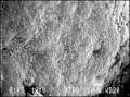
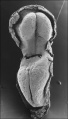
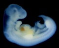
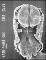
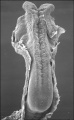
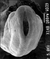
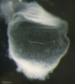
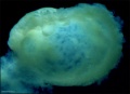
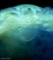
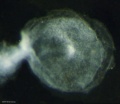
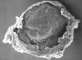
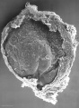
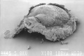
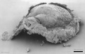
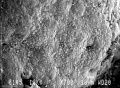
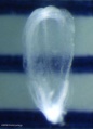
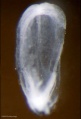
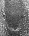
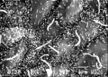
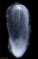
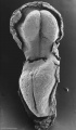
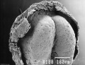
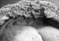
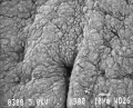
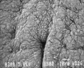
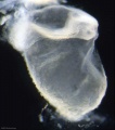
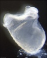
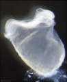
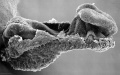
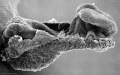
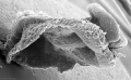
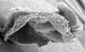
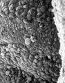
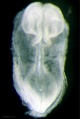
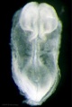
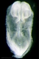
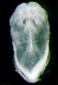
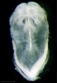
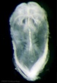
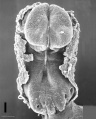
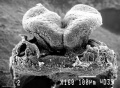
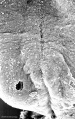
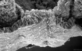
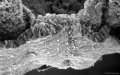
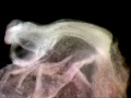
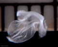
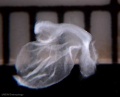
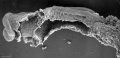
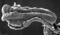
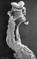
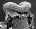
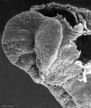
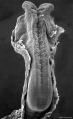
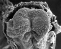
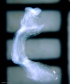
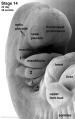
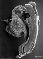
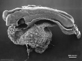
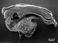
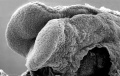
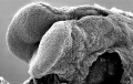
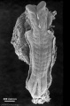
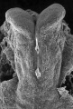
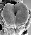
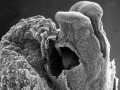
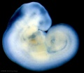
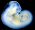
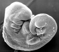
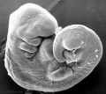
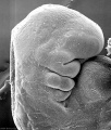
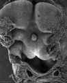
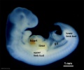
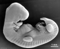
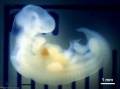
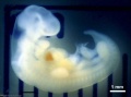
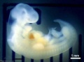
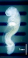
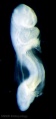
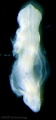
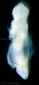
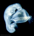
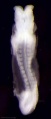
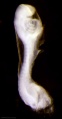
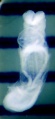
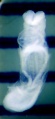
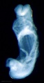
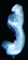
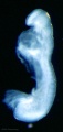
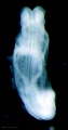
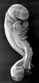
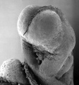
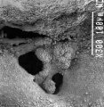
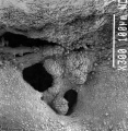
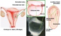
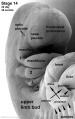
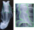
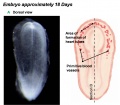
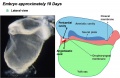
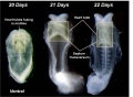
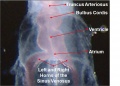
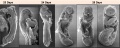
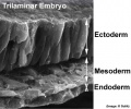
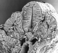
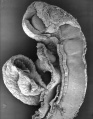
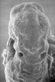
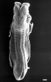
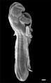
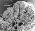
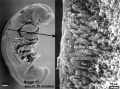
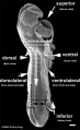
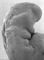
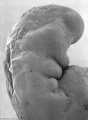
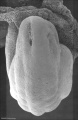
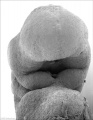
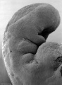
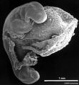
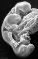
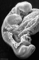
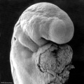
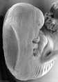
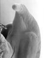
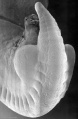
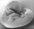
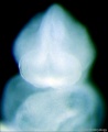
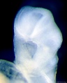
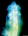
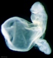
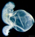
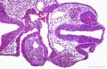
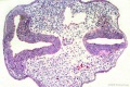
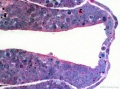
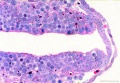
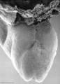
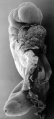
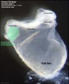
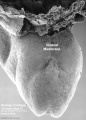
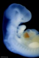
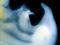
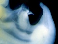
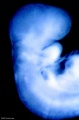
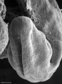
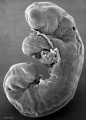
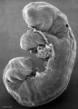
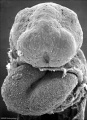
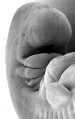
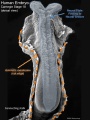
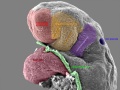
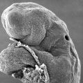
The following images are image excerpts from a collaborative eBook currently in preparation. The images include both brightfieqld and scanning EM images of teh early human embryo between stage 7 to 14 (week 3-5, GA week 5-7).
Image Source: Scanning electron micrographs of the Carnegie stages of the early human embryos are reproduced with the permission of Prof Kathy Sulik, from embryos collected by Dr. Vekemans and Tania Attié-Bitach. Images are for educational purposes only and cannot be reproduced electronically or in writing without permission. ==Gallert

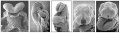


- Carnegie stage 9 gallery


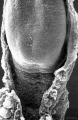


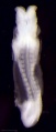


- Stage12 sem7.jpg.jpg


.jpg)
.jpg)


Cite this page: Hill, M.A. (2024, April 26) Embryology Human Embryo SEM. Retrieved from https://embryology.med.unsw.edu.au/embryology/index.php/Human_Embryo_SEM
- © Dr Mark Hill 2024, UNSW Embryology ISBN: 978 0 7334 2609 4 - UNSW CRICOS Provider Code No. 00098G