Histology Stains
From Embryology
Introduction
This page gives a general overview of some histological stains used to identify structures in cells and tissues. This stains information should also be considered in relation to [fixatives].
Medicine Foundations students do not need to know this detail.
Common Stains and Their Reactions | |||||
| Haematoxylin | mucins - light blue | ||||
| Eosin | colloid - pinkmuscle - red | ||||
| Iron Haematoxylin | |||||
| Van Gieson | muscle: yellow/browncartilage - pink | ||||
| Verhoeff's Elastin | elastic fibres - black | ||||
| Tartrazine | |||||
| Silver Impregnation | reticular fibres - black | ||||
| Methyl Green | |||||
| Nuclear Fast Red | |||||
| Gomori's Trichrome | keratin - redmuscle - purple/red | ||||
| Heidenhain's Azan | muscle - red | ||||
| Osmium tetroxide | myelin, lipids - black | ||||
| Alcian Blue | mucins, - blue | ||||
| Periodic acid-Schiff's reagent (PAS) | mucins, glycogen, glycocalyx - magenta | ||||
| PTAH | muscle bands - blue | ||||
| Masson's Trichrome | cartilage, mucins - blue or green; muscle - red | ||||
| Luxol Fast Blue | myelin - blue~ | ||||
| Aldehyde Fuchsin | elastic fibres, mast cells - deep purple | ||||
| Light Green | |||||
| Gallocyanin | nucleic acids, Nissl granules - dark blue | ||||
| Romanowsky(e.g. Leishman's stain) | acidophils - redbasophils - blueazurophilic - purple | ||||
| Aldehyde Pararosanilin | elastic fibres - purple | ||||
Supporting Stain Information
Hematoxylin and Eosin
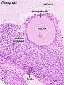
Ovary Histology

Ovary Histology
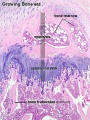
Endochondral ossification


Hematoxylin
- Stains nuclei blue to dark-blue.
- Stains the matrix of hyaline cartilage, myxomatous, and mucoid material pale blue.
- Stains myelin weakly but is not noticeable if combined with eosin stain.
Eosin
- Stains cytoplasm pink to red; red blood cells are also bright red.
- Common counterstain to hematoxylin.
- Stain intensity varies with the formula as well as the fixative.
Periodic acid-Schiff (PAS)
- Stains glycogen, mucin, fungus, basement membrane and other substances.
- Stain used to detect fungal organisms and cytoplasmic accumulation of glycogen.
- Stains lysosomes granules red-purple, can be used in recognition of macrophages.
Alcian Blue
- Stains mucopolysaccharides or glycosaminoglycans
- cationic dye (positively charged molecule) for the demonstration of glycosaminoglycans.
- binds anionic (negative) sites on the polysaccharide.
- can be combined with H&E and VG staining methods.
Masson’s Trichrome Stain
- Stains nuclei deep blue, skeletal and smooth muscles red, collagen and mucin blue.
- Stains brain and spinal cord parenchymal tissue dusky pink to red.
- Used to evaluate fibrosis
- Striations in skeletal muscles also shows up much better in Masson’s trichrome than in hematoxylin and eosin stain.
- Although called a trichrome, four dyes (hematoxylin, Biebrich scarlet, acid fuchsin, and analine blue) are utilized.
PhosphoTungstic Acid Hematoxylin (PTAH)
- Stains nucleus and cytoplasm detail and connective tissue fibers.
- Stains collagen pink, fibrin blue, and striated muscle blue.
- Historic stain used to show CNS reactive astrocytes now used immunochemistry for glial fibrillary acidic protein (GFAP).
Toulidine Blue
- Stains nucleus blue and cytoplasm light blue.
- A synthetic dye in the thiazins family.
Verhoeff-Van Gieson
- Verhoeff-Van Gieson or elastic-Van Gieson (EVG) stain.
- This is a combination of Verhoeff’s elastic stain which is a hematoxylin stain containing ferric chloride and Wright’s iodine solution and Van Gieson stain which contains acid fuchsin, picric acid, and hematoxylin.
- Stains elastic fibers blue-black to black, collagen pale red, other tissue elements yellow, and nuclei blue to black.
Some text modified from: Theory and practice of histological techniques. 3rd edt. By Bancroft JD and Stevens A. Churchill Livingstone, 1990.