File:Wyburn1939-text-fig08-10.jpg: Difference between revisions
From Embryology
mNo edit summary |
mNo edit summary |
||
| Line 1: | Line 1: | ||
{| | |||
| '''Text-fig. 8.''' Embryo 16.1 mm. Reconstruction of a median sagittal section. x 13. Description in text. Vertical lines = liver. Horizontal lines = blood vessels. Black = mesoderm. U .C. = umbilical cord coelom. | |||
| '''Text-fig. 9.''' Embryo 23 mm. Reconstruction of a median sagittal section. x 13. Description in text. Vertical lines = liver. Horizontal lines = blood vessels. Black = mesoderm. U .C. = umbilical cord coelom. | |||
| '''Text-fig. 10.''' Embryo 40 mm. Reconstruction of a median sagittal section. x 10. Description in text. Vertical lines = liver. Horizontal lines = blood vessels. Black = mesoderm. U .C. = umbilical cord coelom. | |||
|} | |||
{{Wyburn1939 figures}} | {{Wyburn1939 figures}} | ||
Latest revision as of 13:34, 15 September 2015
| Text-fig. 8. Embryo 16.1 mm. Reconstruction of a median sagittal section. x 13. Description in text. Vertical lines = liver. Horizontal lines = blood vessels. Black = mesoderm. U .C. = umbilical cord coelom. | Text-fig. 9. Embryo 23 mm. Reconstruction of a median sagittal section. x 13. Description in text. Vertical lines = liver. Horizontal lines = blood vessels. Black = mesoderm. U .C. = umbilical cord coelom. | Text-fig. 10. Embryo 40 mm. Reconstruction of a median sagittal section. x 10. Description in text. Vertical lines = liver. Horizontal lines = blood vessels. Black = mesoderm. U .C. = umbilical cord coelom. |
| Historic Disclaimer - information about historic embryology pages |
|---|
 |
- Links: Plate 1 | Plate 2 | Plate 3 | Plate 4 | Fig 13 | Fig 14 | Fig 15 | Fig 16 | Plate 5 | Fig 17 | Fig 18 | Fig 19 | Fig 20 | Wyburn 1939 | Historic Embryology Papers
Reference
Wyburn GM. The formation of the umbilical cord and the umbilical region of the anterior abdominal wall. (1939) J Anat. 73(2): 289-310.9. PMID 17104757
Cite this page: Hill, M.A. (2024, May 21) Embryology Wyburn1939-text-fig08-10.jpg. Retrieved from https://embryology.med.unsw.edu.au/embryology/index.php/File:Wyburn1939-text-fig08-10.jpg
- © Dr Mark Hill 2024, UNSW Embryology ISBN: 978 0 7334 2609 4 - UNSW CRICOS Provider Code No. 00098G
File history
Click on a date/time to view the file as it appeared at that time.
| Date/Time | Thumbnail | Dimensions | User | Comment | |
|---|---|---|---|---|---|
| current | 13:32, 15 September 2015 | 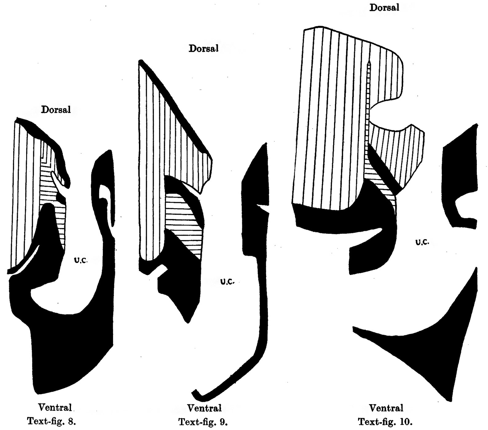 | 1,588 × 1,400 (183 KB) | Z8600021 (talk | contribs) | |
| 13:32, 15 September 2015 | 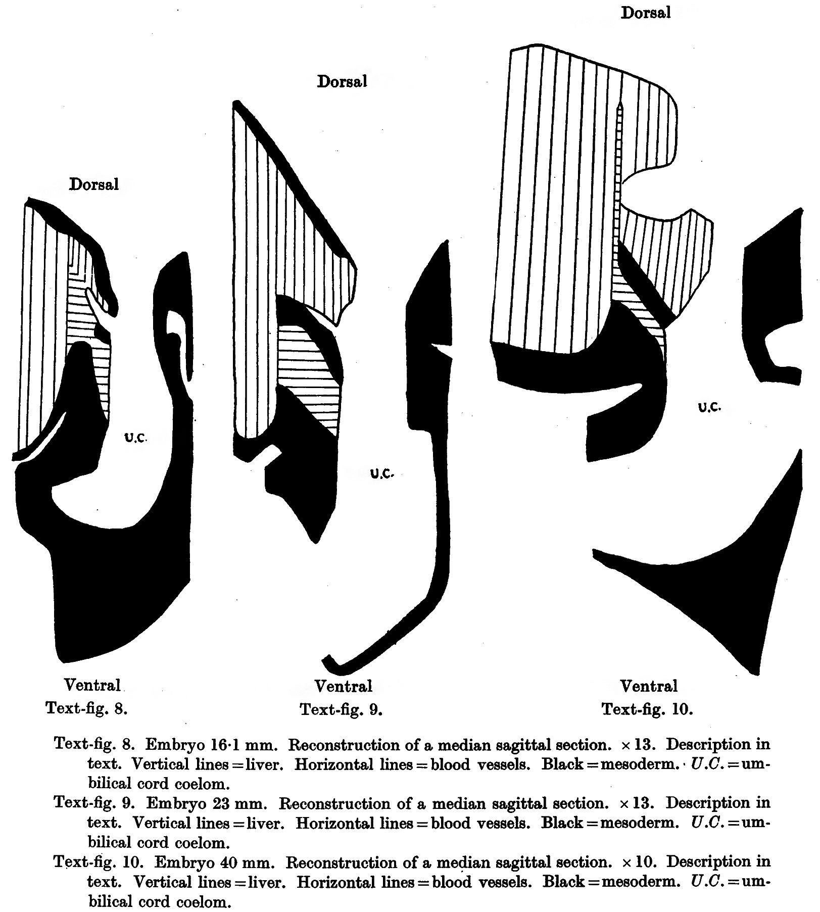 | 1,623 × 1,805 (343 KB) | Z8600021 (talk | contribs) | ||
| 13:30, 15 September 2015 |  | 1,623 × 1,805 (343 KB) | Z8600021 (talk | contribs) |
You cannot overwrite this file.
File usage
The following page uses this file: