File:Wyburn1937 text-fig05.jpg

Original file (1,122 × 545 pixels, file size: 85 KB, MIME type: image/jpeg)
Text-fig. 5. Embryo 12.5 mm
Description in text.
A. Reconstruction of ventral surface from septum transversum to cloacal membrane. x circa 12.
B. Reconstruction of median sagittal section of same. x 10.
Dense mesoderm — black; ventral cloaca and allantois — large dots; cloacal membrane — horizontal lines; blood vessels — oblique lines; liver — circles. A = section 675 at the level of the cranial attachment of body stalk; B = section 775; C = section 825; interval between B and C is the extension of the coelom into the stalk and occupied by mid-gut loop; D = section 875 at the level of caudal attachment of the stalk; GU. = section 888 at the level of the genital tubercle.
File history
Click on a date/time to view the file as it appeared at that time.
| Date/Time | Thumbnail | Dimensions | User | Comment | |
|---|---|---|---|---|---|
| current | 17:33, 15 August 2015 | | 1,122 × 545 (85 KB) | Z8600021 (talk | contribs) | |
| 17:32, 15 August 2015 |  | 1,122 × 545 (85 KB) | Z8600021 (talk | contribs) | ||
| 17:31, 15 August 2015 | 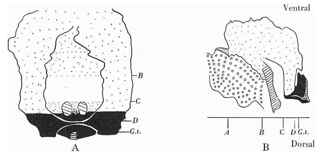 | 1,122 × 545 (85 KB) | Z8600021 (talk | contribs) | ||
| 17:28, 15 August 2015 |  | 1,122 × 545 (85 KB) | Z8600021 (talk | contribs) | ||
| 17:25, 15 August 2015 |  | 1,437 × 879 (235 KB) | Z8600021 (talk | contribs) |
You cannot overwrite this file.
File usage
The following page uses this file: