File:Windle1940 fig03.jpg

Original file (1,000 × 1,077 pixels, file size: 239 KB, MIME type: image/jpeg)
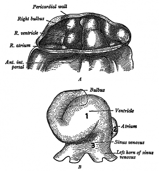
Fig. 3. The heart of human embryos
(A) 6 sornites and (B) 11 somites.
The heart begins to beat in mamrnalian embryos eornparable with L. The order of initiation of the beat is shown in B by the figures 1 (ventricle) , 2 (atriurn) and 3 (sinus venosus).
Image - Arey: "Developmental Anatomy."
1940 Physiology of the Fetus: 1 Introduction | 2 Heart | 3 Circulation | 4 Blood | 5 Respiration | 6 Respiratory Movements | 7 Digestive | 8 Renal - Skin | 9 Muscles | 10 Neural Genesis | 11 Neural Activity | 12 Motor Reactions and Reflexes | 13 Senses | 14 Endocrine | 15 Nutrition and Metabolism | Figures
Reference
Windle WF. Physiology of the Fetus. (1940) Saunders, Philadelphia.
Cite this page: Hill, M.A. (2024, April 27) Embryology Windle1940 fig03.jpg. Retrieved from https://embryology.med.unsw.edu.au/embryology/index.php/File:Windle1940_fig03.jpg
- © Dr Mark Hill 2024, UNSW Embryology ISBN: 978 0 7334 2609 4 - UNSW CRICOS Provider Code No. 00098G
File history
Click on a date/time to view the file as it appeared at that time.
| Date/Time | Thumbnail | Dimensions | User | Comment | |
|---|---|---|---|---|---|
| current | 16:06, 11 September 2018 |  | 1,000 × 1,077 (239 KB) | Z8600021 (talk | contribs) | |
| 16:03, 11 September 2018 |  | 1,351 × 1,394 (398 KB) | Z8600021 (talk | contribs) | {{Windle1940 TOC}} ===Reference=== {{Ref-Windle1940}} {{Footer}} |
You cannot overwrite this file.
File usage
The following page uses this file: