File:Whillis1940 plate02.jpg: Difference between revisions
No edit summary |
(Z8600021 uploaded a new version of File:Whillis1940 plate02.jpg) |
||
| (One intermediate revision by the same user not shown) | |||
| Line 1: | Line 1: | ||
==Plate II== | |||
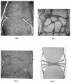
Fig. 6. Interphalangeal joint at 125 mm. stage. x60. 11., Liquefaction of matrix in marginal part of bond of union. | |||
Fig. 6. Wrist region, 30 mm. stage. x 55. a, joint disk between ulnar styloid and triquetral. | |||
Fig. 7. Wrist region, 125 mm. stage. x 20. | |||
Fig. 8. Model illustrating elfect of pressure on a laminated disk formed of sheets of rubber. | |||
===Reference=== | |||
{{Ref-Whillis1940}} | |||
{{Footer}} | |||
[[Category:Human]][[Category:Joint]][[Category:Historic Embryology]][[Category:1940's]] | |||
Latest revision as of 18:43, 16 August 2017
Plate II
Fig. 6. Interphalangeal joint at 125 mm. stage. x60. 11., Liquefaction of matrix in marginal part of bond of union.
Fig. 6. Wrist region, 30 mm. stage. x 55. a, joint disk between ulnar styloid and triquetral.
Fig. 7. Wrist region, 125 mm. stage. x 20.
Fig. 8. Model illustrating elfect of pressure on a laminated disk formed of sheets of rubber.
Reference
Whillis J. The development of synovial joints. (1940) J Anat. 74(Pt 2): 277-283. PMID: 17104813
Cite this page: Hill, M.A. (2024, May 21) Embryology Whillis1940 plate02.jpg. Retrieved from https://embryology.med.unsw.edu.au/embryology/index.php/File:Whillis1940_plate02.jpg
- © Dr Mark Hill 2024, UNSW Embryology ISBN: 978 0 7334 2609 4 - UNSW CRICOS Provider Code No. 00098G
File history
Click on a date/time to view the file as it appeared at that time.
| Date/Time | Thumbnail | Dimensions | User | Comment | |
|---|---|---|---|---|---|
| current | 18:43, 16 August 2017 |  | 1,280 × 1,472 (354 KB) | Z8600021 (talk | contribs) | |
| 18:42, 16 August 2017 |  | 1,887 × 2,509 (912 KB) | Z8600021 (talk | contribs) |
You cannot overwrite this file.
File usage
The following page uses this file: