File:Wheeler1920 fig05-11.jpg

Original file (802 × 1,000 pixels, file size: 168 KB, MIME type: image/jpeg)
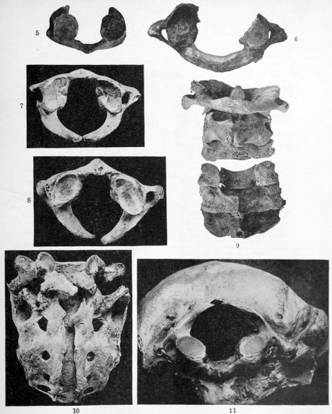
Figs. 5 and 6. Two specimens of the atlas in which there is incomplete dorsal arch formation.
Fig. 5, specimen No. 25S637 F2, U. S. National Museum.
Fig. 6, specimen No. 262993, U. S. National Museum.
Fig. 7. Specimen showing symmetry of the dorsal laminse. Specimen No. 256457, U. S. National Museum.
Fig. 8. Specimen showing asymmetry of the dorsal laminje. Specimen No. 271782, U. S. National Museum.
Fig. 9. Posterior view of the cerv'ical vertebRe, of wliich the fifth lacks dorsal laminae. Specimen No. 227471, U. S. National Museum.
Fig. 10. Specimen showing open Siicral canal and partial sacralization of fifth lumbar vertebra, the dorsal arch of which is bifid. X 0.7. Specimen No. 17980506, U. S. National Museum.
Fig. 11. Specimen showing fusion of atlas and occiput, the posterior arch of the atlas being incomplete.
File history
Click on a date/time to view the file as it appeared at that time.
| Date/Time | Thumbnail | Dimensions | User | Comment | |
|---|---|---|---|---|---|
| current | 23:25, 23 April 2012 |  | 802 × 1,000 (168 KB) | Z8600021 (talk | contribs) |
You cannot overwrite this file.
File usage
The following page uses this file: