File:West1952 plate02.jpg

Original file (1,280 × 1,651 pixels, file size: 284 KB, MIME type: image/jpeg)
Plate 2
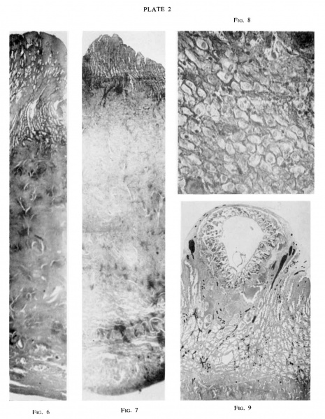
Figs. 6 and 7. Section through the whole thickness of the uterine wall of “ Mal” (6) and
The difference in thickness x 6
Decidual cells, from the upper right corner of fig. 6.
“ Gar” (7) at some distance from the implantation site. of the endometrium in the two specimens is well seen.
“ Mal.” X 385
“ Gar.” The same section as Plate 1, fig. 3. All the vessels have been marked with Indian ink. Many coiled spiral arteries are seen. Larger vessels nearer the ovum are veins. x 10
Reference
West CM. Two presomite human embryos. (1952) J. of Gynaecol. Geat Brit. Emp., 59: 336-351.
Cite this page: Hill, M.A. (2024, April 27) Embryology West1952 plate02.jpg. Retrieved from https://embryology.med.unsw.edu.au/embryology/index.php/File:West1952_plate02.jpg
- © Dr Mark Hill 2024, UNSW Embryology ISBN: 978 0 7334 2609 4 - UNSW CRICOS Provider Code No. 00098G
File history
Click on a date/time to view the file as it appeared at that time.
| Date/Time | Thumbnail | Dimensions | User | Comment | |
|---|---|---|---|---|---|
| current | 13:55, 11 August 2017 |  | 1,280 × 1,651 (284 KB) | Z8600021 (talk | contribs) | |
| 13:54, 11 August 2017 |  | 1,820 × 2,347 (478 KB) | Z8600021 (talk | contribs) |
You cannot overwrite this file.
File usage
The following page uses this file: