File:Watson1915 Fig06.jpg
Original file (1,402 × 1,000 pixels, file size: 226 KB, MIME type: image/jpeg)
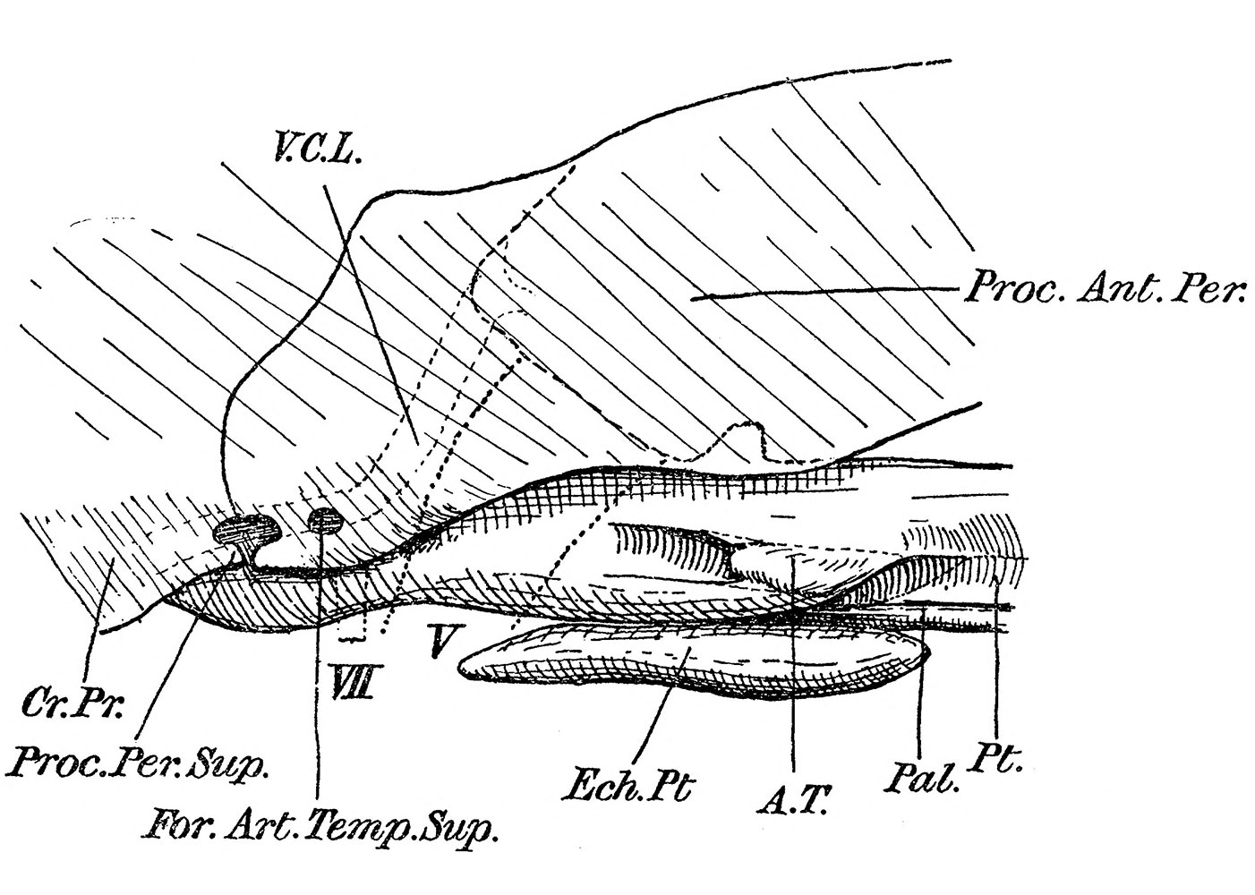
Fig. 6. Reconstruction of the skull of Embryo beta otic and temporal region viewed from the side
Omithorhynchus paradoxus. Graphical reconstruction of the skull of Embryo beta in the otic and temporal region, seen from the side. Reference letters as before, with — For, Art. Temp, Sup., foramen for the superficial temporal artery: Proc.Per.Sup., processus perioticus superior; Froc.AnLPer,, processus anterior perioticus, the ossification in the membrana spheno-obturatoria which belongs to the prootic. The broken line lying within this bone represents the front edge of the cartilaginous otic capsule : V,C,L., vena capitis lateralis, course in the sulcus facialis and the canal in the prootic; VII, the position of the facial foramen ; F, the mandibular division of the fifth nerve.
- The Monotreme Skull: Fig 1 | Fig 2 | Fig 3 | Fig 4 | Fig 5 | Fig 6 | Fig 7 | Fig 8 | Fig 9 | Fig 10 | Fig 11 | Fig 12 | Fig 13 | Fig 14 | Fig 15 | Fig 16 | Fig 17 | Fig 18 | Fig 19 | Plate 1 | Plate 2 | Plate 3 | Plate 4
| Historic Disclaimer - information about historic embryology pages |
|---|
 |
Reference
Watson DMS. The Monotreme Skull - A Contribution to Mammalian Morphogenesis. (1915)
Cite this page: Hill, M.A. (2024, April 27) Embryology Watson1915 Fig06.jpg. Retrieved from https://embryology.med.unsw.edu.au/embryology/index.php/File:Watson1915_Fig06.jpg
- © Dr Mark Hill 2024, UNSW Embryology ISBN: 978 0 7334 2609 4 - UNSW CRICOS Provider Code No. 00098G
File history
Click on a date/time to view the file as it appeared at that time.
| Date/Time | Thumbnail | Dimensions | User | Comment | |
|---|---|---|---|---|---|
| current | 22:50, 30 October 2013 | | 1,402 × 1,000 (226 KB) | Z8600021 (talk | contribs) | ==Fig. 6.== The same model fig. 1., {{Watson1915 figures}} |
You cannot overwrite this file.
File usage
The following page uses this file: