File:Waterston1915 fig03.jpg: Difference between revisions
(Z8600021 uploaded a new version of File:Waterston1915 fig03.jpg) |
mNo edit summary |
||
| Line 1: | Line 1: | ||
==Fig. 3. Dorsal wall and floor of pericardial sac of 22 mm Embryo== | |||
A, right duct of Cuvier; B, aacendin aorta: C, dorsal arterial mesocardium ; D, arterial mesocardium; E, left duct of uvier, divided; F, meuocardial told; G vena cava Interior; H, floor of pericardium; K. pulmonary veins; L, pleura and pericardial wall; M, apex of lung; N, pleural cavity: P, pulmonary artery. | |||
{{Waterston1915 figures}} | {{Waterston1915 figures}} | ||
Revision as of 16:46, 24 August 2015
Fig. 3. Dorsal wall and floor of pericardial sac of 22 mm Embryo
A, right duct of Cuvier; B, aacendin aorta: C, dorsal arterial mesocardium ; D, arterial mesocardium; E, left duct of uvier, divided; F, meuocardial told; G vena cava Interior; H, floor of pericardium; K. pulmonary veins; L, pleura and pericardial wall; M, apex of lung; N, pleural cavity: P, pulmonary artery.
| Historic Disclaimer - information about historic embryology pages |
|---|
 |
Reference
Waterston D. Developmental changes in the pericardium, the mesocardia, and the pleural sacs in the human embryo. (1915) J Anat Physiol., 50(1): 24-9. PMID 17233049
Cite this page: Hill, M.A. (2024, April 27) Embryology Waterston1915 fig03.jpg. Retrieved from https://embryology.med.unsw.edu.au/embryology/index.php/File:Waterston1915_fig03.jpg
- © Dr Mark Hill 2024, UNSW Embryology ISBN: 978 0 7334 2609 4 - UNSW CRICOS Provider Code No. 00098G
File history
Click on a date/time to view the file as it appeared at that time.
| Date/Time | Thumbnail | Dimensions | User | Comment | |
|---|---|---|---|---|---|
| current | 16:46, 24 August 2015 |  | 767 × 937 (153 KB) | Z8600021 (talk | contribs) | |
| 16:45, 24 August 2015 | 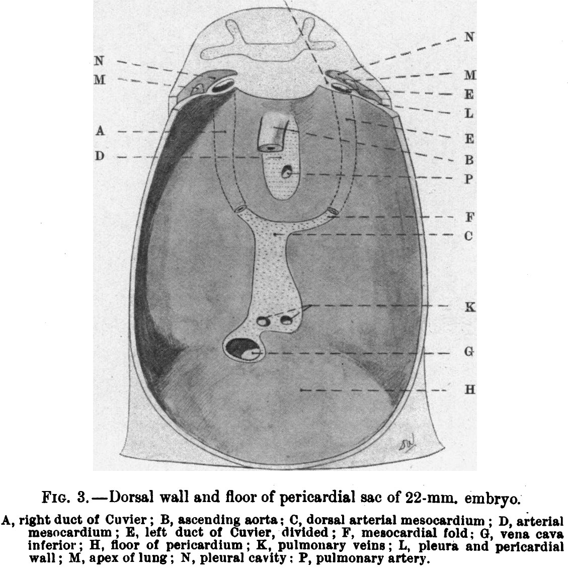 | 1,129 × 1,135 (269 KB) | Z8600021 (talk | contribs) | {{Waterston1915 figures}} |
You cannot overwrite this file.
File usage
The following page uses this file: