File:Wallaby embryo 04.jpg
Original file (1,138 × 1,098 pixels, file size: 110 KB, MIME type: image/jpeg)
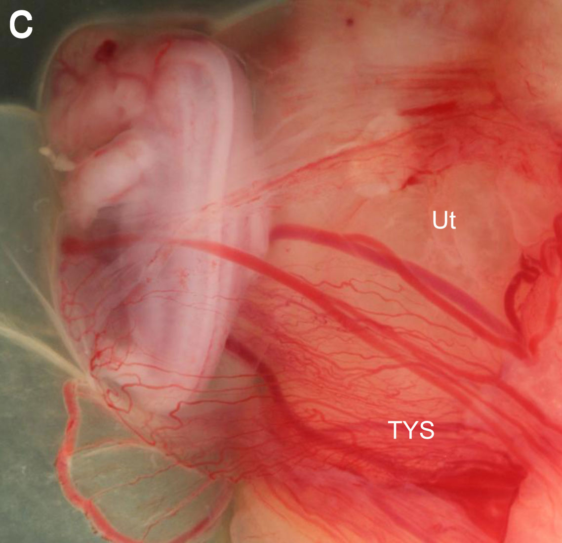
Tammar Wallaby Embryo and Fetus
Fetus at day 23 of pregnancy, showing the increase in the vascular region and the close attachment of the placenta to the uterine epithelium (Ut). The vitelline vessels are prominent.
Legend
- All - allantois
- Am - amniopore
- BYS - bilaminar yolk sac
- TYS - trilaminar yolk sac
- Sh - shell coat
- ST - sinus terminalis
- Ut - uterine epithelium
- Wallaby Links: Wallaby Embryo and Fetus | Embryo and Membranes day 18 | Embryo day 18 | Fetus day 23 | Fetus day 25 | Kangaroo Development
Reference
<pubmed>21854600</pubmed>| PMC3170617 | Evodevo.
Menzies et al. EvoDevo 2011 2:16 doi:10.1186/2041-9139-2-16
© 2011 Menzies et al; licensee BioMed Central Ltd.
This is an Open Access article distributed under the terms of the Creative Commons Attribution License (http://creativecommons.org/licenses/by/2.0), which permits unrestricted use, distribution, and reproduction in any medium, provided the original work is properly cited.
File history
Click on a date/time to view the file as it appeared at that time.
| Date/Time | Thumbnail | Dimensions | User | Comment | |
|---|---|---|---|---|---|
| current | 13:11, 23 May 2012 | | 1,138 × 1,098 (110 KB) | Z8600021 (talk | contribs) | ==Tammar Wallaby Embryo and Fetus== (a) At day 18, the conceptus emerges form the ruptured shell coat (Sh) so that the two regions of the placenta can make a close attachment to the uterine epithelium. The right side of the embryo is already coat-free, w |
You cannot overwrite this file.
File usage
The following page uses this file: