File:Vomeronasal Organ position.jpg: Difference between revisions
From Embryology
No edit summary |
No edit summary |
||
| Line 2: | Line 2: | ||
This image demonstrates the position of the vomeronasal organ in relation to the olfactory bulb and tract. | This image demonstrates the position of the vomeronasal organ in relation to the olfactory bulb and tract. | ||
--[[User:Z8600021|Mark Hill]] 00:41, 3 October 2012 (EST) More supporting information could be supplied here. What is this image based upon? | |||
{{Template:Student Image}} | {{Template:Student Image}} | ||
Revision as of 00:41, 3 October 2012
Student drawn image
This image demonstrates the position of the vomeronasal organ in relation to the olfactory bulb and tract.
--Mark Hill 00:41, 3 October 2012 (EST) More supporting information could be supplied here. What is this image based upon?
- Note - This image was originally uploaded as part of an undergraduate science student project and may contain inaccuracies in either description or acknowledgements. Students have been advised in writing concerning the reuse of content and may accidentally have misunderstood the original terms of use. If image reuse on this non-commercial educational site infringes your existing copyright, please contact the site editor for immediate removal.
File history
Click on a date/time to view the file as it appeared at that time.
| Date/Time | Thumbnail | Dimensions | User | Comment | |
|---|---|---|---|---|---|
| current | 13:50, 1 October 2012 | 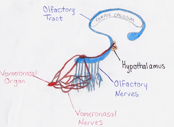 | 565 × 413 (49 KB) | Z3374215 (talk | contribs) | Student drawn image This image demonstrates the position of the vomeronasal organ in relation to the olfactory bulb and tract. {{Template:Student Image}} |
You cannot overwrite this file.
File usage
The following 2 pages use this file: