File:Ultrasound - fetal abdominal circumference.jpg
Ultrasound_-_fetal_abdominal_circumference.jpg (672 × 512 pixels, file size: 23 KB, MIME type: image/jpeg)
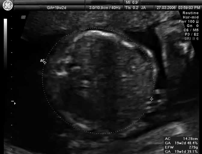
Human Fetal Ultrasound (GA 19 weeks)
Ultrasound (GA19 weeks) scan through the fetal trunk to measure abdominal circumference (AC).
An ultrasound measurement of Abdominal Circumference (AC) is used to determine fetal age and normal development (small/large/abnormal) parameters. Measured at the outer edge of the circumference of the body plane in which the portal vein or stomach can be visualized in a tangential section. It is one of the four typical ultrasound assessments of fetal size and age: Biparietal Diameter (BPD), Head Circumference (HC), Abdominal Circumference (AC), and Femur Length (FL).
Abdominal Circumference of less than 31 cm at 36 to 40 weeks gestation is a predictor of intrauterine growth retardation (IUGR).
- Links: Ultrasound | Fetal Development | Musculoskeletal System
Cite this page: Hill, M.A. (2024, April 27) Embryology Ultrasound - fetal abdominal circumference.jpg. Retrieved from https://embryology.med.unsw.edu.au/embryology/index.php/File:Ultrasound_-_fetal_abdominal_circumference.jpg
- © Dr Mark Hill 2024, UNSW Embryology ISBN: 978 0 7334 2609 4 - UNSW CRICOS Provider Code No. 00098G
File history
Click on a date/time to view the file as it appeared at that time.
| Date/Time | Thumbnail | Dimensions | User | Comment | |
|---|---|---|---|---|---|
| current | 12:34, 30 May 2010 | | 672 × 512 (23 KB) | S8600021 (talk | contribs) | Ultrasound Fetal (GA 19 weeks) Ultrasound scan through the fetal trunk to measure abdominal circumference (AC). Category:Ultrasound Category:Fetal Category:Second Trimester |
You cannot overwrite this file.