File:Ultrasound12wk 3D image.jpg: Difference between revisions
From Embryology
No edit summary |
No edit summary |
||
| Line 1: | Line 1: | ||
A 3D ultrasound static image of the 12 week fetus shows a ventral view with the fetus upside down, with the head down and cord to the top. | A 3D ultrasound static image of the 12 week fetus shows a ventral view with the fetus upside down, with the head down and cord to the top. | ||
Scale bar 1 cm | |||
Original file name: 12wk2_3D2.jpg | Original file name: 12wk2_3D2.jpg | ||
Latest revision as of 15:10, 11 October 2009
A 3D ultrasound static image of the 12 week fetus shows a ventral view with the fetus upside down, with the head down and cord to the top.
Scale bar 1 cm
Original file name: 12wk2_3D2.jpg
Image source: UNSW Embryology http://embryology.med.unsw.edu.au/Movies/usound/Hum3D.htm
File history
Click on a date/time to view the file as it appeared at that time.
| Date/Time | Thumbnail | Dimensions | User | Comment | |
|---|---|---|---|---|---|
| current | 16:44, 5 August 2009 | 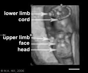 | 301 × 248 (8 KB) | S8600021 (talk | contribs) | A 3D ultrasound static image of the 12 week fetus shows a ventral view with the fetus upside down, with the head down and cord to the top. |
You cannot overwrite this file.