File:Trophoblast HLA-G expression 01.jpg
Original file (791 × 688 pixels, file size: 146 KB, MIME type: image/jpeg)
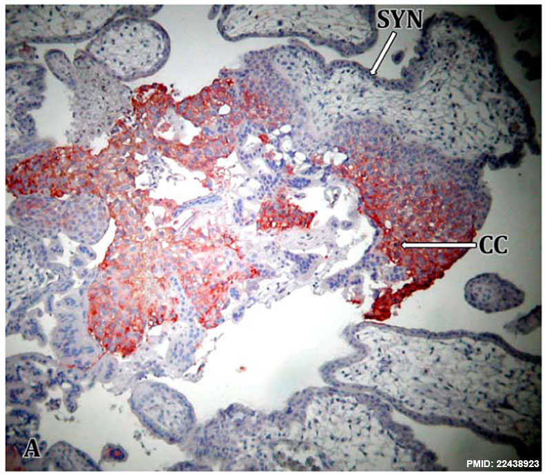
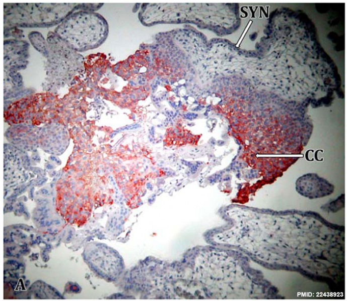
Trophoblast HLA-G expression
Immunohistochemisty performed on first trimester placental tissue section.
- HLA-G expression (red) is detected in the trophoblast cell columns (CC) and extravillous trophoblasts.
- HLA-G expression staining not seen for syncitiotrophoblast (SYN) cells.
Reference
<pubmed>22438923</pubmed>| PLoS One.
Copyright
© 2012 Manaster et al. This is an open-access article distributed under the terms of the Creative Commons Attribution License, which permits unrestricted use, distribution, and reproduction in any medium, provided the original author and source are credited.
Manaster I, Goldman-Wohl D, Greenfield C, Nachmani D, Tsukerman P, et al. (2012) MiRNA-Mediated Control of HLA-G Expression and Function. PLoS ONE 7(3): e33395. doi:10.1371/journal.pone.0033395
Figure 1. doi:10.1371/journal.pone.0033395.g001 Journal.pone.0033395.g001.jpg
Right panel A cropped, resized and relabeled from Figure 1.
File history
Click on a date/time to view the file as it appeared at that time.
| Date/Time | Thumbnail | Dimensions | User | Comment | |
|---|---|---|---|---|---|
| current | 12:16, 17 March 2014 | | 791 × 688 (146 KB) | Z8600021 (talk | contribs) | ==Trophoblast HLA-G expression== Figure 1. MiR-148a and miR-152 potentially target the 3′UTR of HLA-G. (A) Immunohistochemisty performed on first trimester placental tissue sections. Right panel: negative control. Left panel: HLA-G expression is de... |
You cannot overwrite this file.
File usage
The following page uses this file: