File:Trisomy X.jpg
Trisomy_X.jpg (400 × 352 pixels, file size: 15 KB, MIME type: image/jpeg)
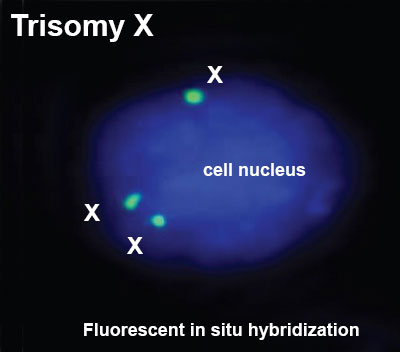
Trisomy X
Trisomy X, 3 copies of the X chromosome identified in a single cell nucleus by fluorescent in situ hybridisation (FISH) analysis.
- Links: Trisomy X | Abnormal Development - Genetic |
Reference
Venkateshwari A, Srimanjari K, Srilekha A, Begum A, Sujatha M, Sunitha T, Nallari P & Jyothy A. (2012). Mosaic triple X syndrome in a female with primary amenorrhea. Indian J Hum Genet , 18, 246-9. PMID: 23162306 DOI.
Copyright
Original Figure 2 cropped, resized and labelled. Indian J HumGenet_2012_18_2_246_100790_u2.jpg
Cite this page: Hill, M.A. (2024, April 27) Embryology Trisomy X.jpg. Retrieved from https://embryology.med.unsw.edu.au/embryology/index.php/File:Trisomy_X.jpg
- © Dr Mark Hill 2024, UNSW Embryology ISBN: 978 0 7334 2609 4 - UNSW CRICOS Provider Code No. 00098G
File history
Click on a date/time to view the file as it appeared at that time.
| Date/Time | Thumbnail | Dimensions | User | Comment | |
|---|---|---|---|---|---|
| current | 20:34, 24 December 2013 |  | 400 × 352 (15 KB) | Z8600021 (talk | contribs) | ==Trisomy X== Trisomy X, 3 copies of the X chromosome identified in a single cell nucleus by fluorescent in situ hybridisation (FISH) analysis. ===Reference=== <pubmed>23162306</pubmed>| [http://www.ijhg.com/article.asp?issn=0971-6866;year=2012;vol... |
You cannot overwrite this file.
File usage
The following 4 pages use this file:
