File:Tonsil histology 01.jpg: Difference between revisions
No edit summary |
|||
| Line 4: | Line 4: | ||
* the "tonsils", lateral wall of oropharynx | * the "tonsils", lateral wall of oropharynx | ||
* covered by stratified squamous epithelium | * covered by stratified squamous epithelium | ||
* numerous crypts (10-20) infolds of surface epithelium | * numerous crypts (10-20) infolds of surface epithelium | ||
'''Tonsilar Crypt''' (crypt) - palatine tonsil squamous epithelium infold, with intraepithelial passages containing non-epithelial cells. Functions include: [https://www.ncbi.nlm.nih.gov/pubmed/7559106 PMID 7559106] | |||
# intimate contact between immune response effector cells | |||
# facilitate transport of antigens | |||
# synthesise secretory components | |||
# contain a pool of immunoglobulins | |||
* Afferent lymph vessels absent | * Afferent lymph vessels absent | ||
* Efferent lymph vessels are present | * Efferent lymph vessels are present | ||
Latest revision as of 08:10, 18 February 2019
Palatine Tonsils Histology
Anatomical location - Palatine (tonsils), Lingual and Pharyngeal ( adenoids )
- the "tonsils", lateral wall of oropharynx
- covered by stratified squamous epithelium
- numerous crypts (10-20) infolds of surface epithelium
Tonsilar Crypt (crypt) - palatine tonsil squamous epithelium infold, with intraepithelial passages containing non-epithelial cells. Functions include: PMID 7559106
- intimate contact between immune response effector cells
- facilitate transport of antigens
- synthesise secretory components
- contain a pool of immunoglobulins
- Afferent lymph vessels absent
- Efferent lymph vessels are present
- Immune Images: Oesophagus MALT | Colon MALT | Peyer's patch overview | Peyer's patch detail | Cartoon - IEL development | Cartoon - IEL function | Cartoon - IEL differentiation | Mesenteric Lymph Nodes overview | Palatine Tonsil | Tonsil | Immune System Development
Links: Histology | Histology Stains | Blue Histology images copyright Lutz Slomianka 1998-2009. The literary and artistic works on the original Blue Histology website may be reproduced, adapted, published and distributed for non-commercial purposes. See also the page Histology Stains.
Cite this page: Hill, M.A. (2024, May 14) Embryology Tonsil histology 01.jpg. Retrieved from https://embryology.med.unsw.edu.au/embryology/index.php/File:Tonsil_histology_01.jpg
- © Dr Mark Hill 2024, UNSW Embryology ISBN: 978 0 7334 2609 4 - UNSW CRICOS Provider Code No. 00098G
Tns02he.jpg
File history
Click on a date/time to view the file as it appeared at that time.
| Date/Time | Thumbnail | Dimensions | User | Comment | |
|---|---|---|---|---|---|
| current | 09:55, 24 February 2012 | 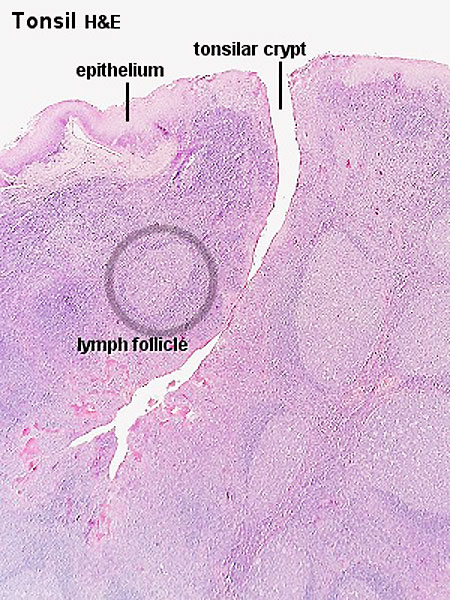 | 450 × 600 (106 KB) | Z8600021 (talk | contribs) | ==Palatine Tonsils Histology== Anatomical location - Palatine (tonsils), Lingual and Pharyngeal ( adenoids ) * the "tonsils", lateral wall of oropharynx * covered by stratified squamous epithelium * numerous crypts (10-20) infolds of surface epithelium |
You cannot overwrite this file.
File usage
The following 3 pages use this file: