File:Thymosin B4 detection in foetal developing ileum.jpg
Original file (427 × 628 pixels, file size: 234 KB, MIME type: image/jpeg)
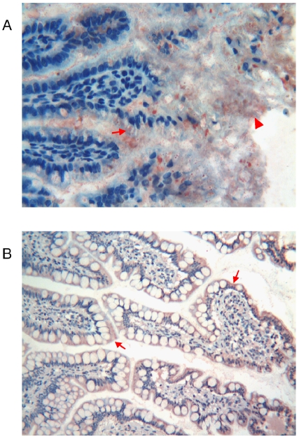
A)Foetal stomach with Tβ4-immunoreactive granules shown by arrow. Arrow head show the Tβ4 granular deposits located in the mucous of the gastric surface.
B)Stomach of adult with intense reactivity for Tβ4 (arrow)
[1].
- ↑ <pubmed>20161756</pubmed>
Copyright
Copyright Nemolato et al. This is an open-access article distributed under the terms of the Creative Commons Attribution License, which permits unrestricted use, distribution, and reproduction in any medium, provided the original author and source are credited.
Note - This image was originally uploaded as part of an undergraduate science student project and may contain inaccuracies in either description or acknowledgements. Students have been advised in writing concerning the reuse of content and may accidentally have misunderstood the original terms of use. If image reuse on this non-commercial educational site infringes your existing copyright, please contact the site editor for immediate removal.
--Mark Hill (talk) 10:21, 7 November 2014 (EST) Assessment - Figure relates to project topic contains reference and copyright. Why was the student template not used here? Minor, formatting is very poor. If Thymosin Beta 4 was to be included in current research it should at least explain the function of the protein in the associated information or on the project page.
File history
Click on a date/time to view the file as it appeared at that time.
| Date/Time | Thumbnail | Dimensions | User | Comment | |
|---|---|---|---|---|---|
| current | 08:53, 23 October 2014 | | 427 × 628 (234 KB) | Z3415242 (talk | contribs) | A)Foetal stomach with Tβ4-immunoreactive granules shown by arrow. Arrow head show the Tβ4 granular deposits located in the mucous of the gastric surface. B)Stomach of adult with intense reactivity for Tβ4 (arrow) <ref name="PMID20161756 "><pubme... |
You cannot overwrite this file.
File usage
The following 2 pages use this file: