File:The morphology of follicles after ovarian tissue vitrification.jpg
The_morphology_of_follicles_after_ovarian_tissue_vitrification.jpg (776 × 165 pixels, file size: 76 KB, MIME type: image/jpeg)
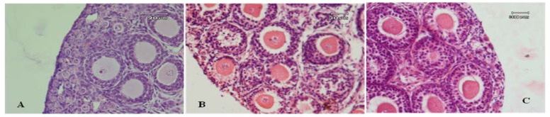
The morphology of follicles after ovarian tissue vitrification
The morphology of follicles after ovarian tissue vitrification: A) non vitrified, B) slow freezing and C) vitrification. No significant differences were observed between the normality of primordial and primary follicles in all groups of study. But slow freezing groups showed more sign of degeneration and cryoinjury in preantral follicles and the disruption of intercellular contacts among innermost granulosa layer and oocyte, nuclear piknosis and cytoplasmic retraction were prominent [1] .
(Original figure legend :IJCP-06-123f2)
References
- ↑ <pubmed>25250122</pubmed>
Copyright
This work is licensed under a Creative Commons Attribution-NonCommercial 3.0 Unported License which allows users to read, copy, distribute and make derivative works for non-commercial purposes from the material, as long as the author of the original work is cited properly.
- Note - This image was originally uploaded as part of an undergraduate science student project and may contain inaccuracies in either description or acknowledgements. Students have been advised in writing concerning the reuse of content and may accidentally have misunderstood the original terms of use. If image reuse on this non-commercial educational site infringes your existing copyright, please contact the site editor for immediate removal.
File history
Click on a date/time to view the file as it appeared at that time.
| Date/Time | Thumbnail | Dimensions | User | Comment | |
|---|---|---|---|---|---|
| current | 16:09, 17 October 2015 | 776 × 165 (76 KB) | Z3463890 (talk | contribs) | (Original figure legend :IJCP-06-123f2) PMID 25250122 |
You cannot overwrite this file.
File usage
The following 2 pages use this file: