File:Thalidomide abnormalities 02.jpg
From Embryology

Size of this preview: 543 × 600 pixels. Other resolution: 724 × 800 pixels.
Original file (724 × 800 pixels, file size: 74 KB, MIME type: image/jpeg)
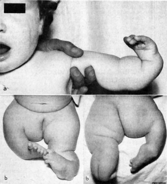
Thalidomide affected Infant
Girl, aged 1 year, whose mother states she had taken thalidomide for 12 months (before and during pregnancy)
- a - The left, arm shows short deformed radius and ulna fused at upper end, and only four metacarpals.
- b - Shows right leg with deformed femur. The upper end is not clearly seen, but fragmented epiphysis is present. fibula is missing. Left leg has head of femur epiphysis missing. The tibia is short and deformed, fibula is missing, and both hips, knees and ankles are dislocated.
- Thalidomide Links: image 1 - limb abnormalities | image 2 - limb abnormalities | Thalidomide | Limb Abnormalities | Limb Development
Reference
GILLIS L. (1962). Thalidomide babies: management of limb defects. Br Med J , 2, 647-51. PMID: 13898662
Fig 3. resized and relabelled.
Cite this page: Hill, M.A. (2024, April 27) Embryology Thalidomide abnormalities 02.jpg. Retrieved from https://embryology.med.unsw.edu.au/embryology/index.php/File:Thalidomide_abnormalities_02.jpg
- © Dr Mark Hill 2024, UNSW Embryology ISBN: 978 0 7334 2609 4 - UNSW CRICOS Provider Code No. 00098G
File history
Click on a date/time to view the file as it appeared at that time.
| Date/Time | Thumbnail | Dimensions | User | Comment | |
|---|---|---|---|---|---|
| current | 10:37, 7 April 2016 |  | 724 × 800 (74 KB) | Z8600021 (talk | contribs) | |
| 10:37, 7 April 2016 |  | 1,000 × 1,286 (179 KB) | Z8600021 (talk | contribs) | ===Reference=== <pubmed>13898662</pubmed> Fig 1. resized and relabelled. {{Footer}} Category:HumanCategory:Abnormal DevelopmentCategory:Limb |
You cannot overwrite this file.
File usage
The following 2 pages use this file: