File:Testis histology.jpg
Testis_histology.jpg (400 × 500 pixels, file size: 54 KB, MIME type: image/jpeg)
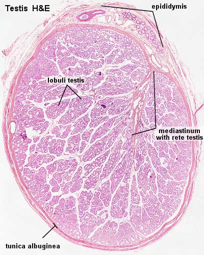
Testis, young and mature - H&E
Use the lowest magnification available. Identify the capsule and the connective tissue septa extending from it. Identify lobules, convoluted seminiferous tubules and clusters of interstitial cells. The mediastinum testis and rete testis are not visible in all sections.
Image Source: UWA Blue Histology http://www.lab.anhb.uwa.edu.au/mb140/CorePages/MaleRepro/malerepro.htm#Testes
Copyright Lutz Slomianka 1998-2009. The literary and artistic works (excluding the UWA logo) on this web-site may be reproduced, adapted, published and distributed for noncommercial purposes. Acknowledgement of the author in any such reproduction, adaption or publication would be appreciated.
Links: Histology | Histology Stains | Blue Histology images copyright Lutz Slomianka 1998-2009. The literary and artistic works on the original Blue Histology website may be reproduced, adapted, published and distributed for non-commercial purposes. See also the page Histology Stains.
Cite this page: Hill, M.A. (2024, May 19) Embryology Testis histology.jpg. Retrieved from https://embryology.med.unsw.edu.au/embryology/index.php/File:Testis_histology.jpg
- © Dr Mark Hill 2024, UNSW Embryology ISBN: 978 0 7334 2609 4 - UNSW CRICOS Provider Code No. 00098G
File history
Click on a date/time to view the file as it appeared at that time.
| Date/Time | Thumbnail | Dimensions | User | Comment | |
|---|---|---|---|---|---|
| current | 16:26, 21 September 2009 | | 400 × 500 (54 KB) | S8600021 (talk | contribs) | |
| 16:25, 21 September 2009 | 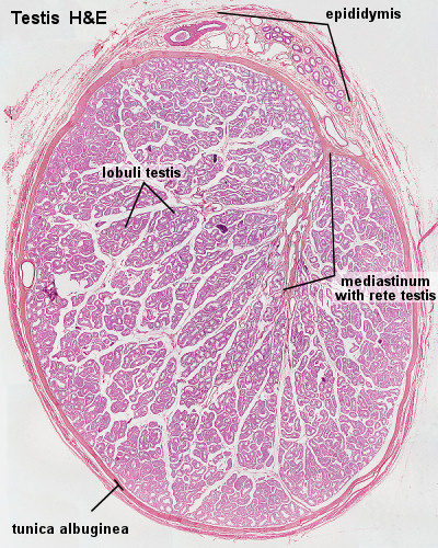 | 400 × 500 (158 KB) | S8600021 (talk | contribs) | Testis, young and mature - H&E Use the lowest magnification available. Identify the capsule and the connective tissue septa extending from it. Identify lobules, convoluted seminiferous tubules and clusters of interstitial cells. The mediastinum testis an |
You cannot overwrite this file.